
Surgery
RLHoLEP Case 35: En bloc Virtual Basket HoLEP 400 grams
Dr. Fernando Gómez Sancha · ICUA
Dr. Fernando Gómez Sancha · YouTube
Language: English. YouTube audio metadata.
Surgical content for professionals. Contains footage of an operation.
YouTube applies an age restriction: if playback is blocked, open the video on YouTube.
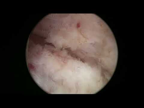
Playback connects to YouTube. Your search query is not sent to the player.
About this video
An en-bloc Virtual Basket HoLEP in a patient in urinary retention, with an estimated prostate size of 400 grams. The description reports a three-hour operation that required division of the prostate at the end and some intracapsular morcellation. It also reports discharge without a catheter the following morning; this outcome belongs to the documented individual case.
Documented details and sources
Only details explicitly stated in the sources. Missing information does not mean it did not occur.
- En bloc
RLHoLEP Case 35: En bloc Virtual Basket HoLEP 400 grams
- Virtual Basket
RLHoLEP Case 35: En bloc Virtual Basket HoLEP 400 grams
- Documented challenging case
This is a recent challenging case, a patient with an estimated size of 400 grams prostate in retention. The operation took 3 hours, but we were able to complete the enucleation and send the pat
- Urinary retention
t challenging case, a patient with an estimated size of 400 grams prostate in retention. The operation took 3 hours, but we were able to complete the enucleation and send the patient home without a catheter the next morning. We had to split the pr
- 400 g
RLHoLEP Case 35: En bloc Virtual Basket HoLEP 400 grams
- Intracapsular morcellation
We had to split the prostate at the end, and do some intracapsular morcellation to be able to finish.
- Splitting the adenoma
We had to split the prostate at the end
Source checked: 2026-10-06.
Available transcript · English
Subtitles exported from the original-language YouTube Studio editor. Not clinically reviewed; source identity and timestamps verified, full audio coverage not verified.
Selected spellings of enucleation, morcellation and holmium terms have been edited. This is not a full audio or clinical review; the original captions remain the source.
They may contain transcription or translation errors; check the explanation in the video.
- hello this is sancho and this is another case of a holmium enucleation of the prostate in a patient with a 400 gram prostate gland it is an extreme case we don't usually see too many patients with this kind of prostate volume but with experience i have seen that it is feasible to operate these larger glands although some things change a little bit in the way we prepare the the patient uh what we tell him and how we perform the operation we are quite convinced that it is possible to nucleate very very large glands carefully in this cases my main concern is to protect the sphincter and of course it takes longer because it is much more surface that we have to dissect and it is much more tissue that
- we have to morsel it so basically when i speak to the patient i tell you know we're going to try to do this endoscopically but there is a chance that we might have to do a second stage or there is a chance that we might need to do a cystotomy to to remove a very large gland also i question them what would you like would you that rather have a small wound below the umbilicus or would you rather have a second stage you see that it's very typical if you force your entry a little bit into the bladder there might be some 12 o'clock split in the mucosa and here i'm marking the the white line just like any other uh case of and blocking nucleation so marking the white line what you will notice is that
- the progress is a little bit slower because we want to pay as we go with hemostasis so i think in this very large glance when you're going to have a very rough and very large surface of dissection it's important to to take good care of hemostasis as you go and even to stop and perform hemostasis if you need to so this is again the white line trying to demarcate trying to incise the mukusa in the interface between adenoma and sphincter trying to have a reference for the rest of the procedure and um that will help protect the sphincter and make make the procedure much safer of course for for the patient so this is the entry into the plane here you can see that i'm moving much slower than usually because i want the
- hemostatic effect of of virtual baskets to to really provide a reliable hemistasis as as we go this is the entry the development of the posterior plane below the apex of the right side of of the prostate you see we have a nice place a nice plane and you see how holmium helps spreading the plane opening the plane and with virtual basket of course and moses we have observed much better hemispheres this is the access now to the to the other side i don't know there are many factors when you do a very large gland like this you have to be extremely calm and you have to be extremely patient because and also you have to trust the method you have to trust the step-by-step approach that you're going to follow
- so if you progress slowly if you carry out carry on with the operation trying to make every movement count every movement when i go to the right when i go to the left i try to to make sure that it is a movement that is going to add uh something to to the procedure so in this case i think it was nearly one hour and a half the the most the nucleation time and then it took us one hour and a half to do the morcellation for some reason most later was not at its peak efficiency uh and it took a little bit while longer than we expected but this patient was then sent home the next morning without a catheter which is a big big big success for such a for such a case so here you can see how i always try to have a line
- that i can follow i do wide movements from side to side usually a little bit faster but in this case as i said it's it's it's paramount to have a good hemistasis and to make sure that the hemostasis will be reliable as you progress so i'm not in a hurry and i progress much more slowly in the larger glands trying to uh get this wonderful hemistasis of course these cases are challenging and i think it's very important to use the the smaller glance to train your ability to perform hollop to train your ability to make every movement count and of course to increase the knowledge about anatomy i have edited the video of course it's not going to be three hours it's going to be a little bit less than an hour
- but i have selected those relevant pieces of video that will hopefully illustrate how one of these prostates can be performed can be done so now we have a posterior line of dissection you see that i try to keep my scope so i can see the line more or less in the middle of the screen so i can get to see a little bit of adenoma and a little bit of capsule and the dissection line is in the middle the line of attack the line of dissection i of course now turn the fiber towards 12 o'clock that's the sphincter with the split that we saw and here i'm trying to certain where where's the limit that's probably the white line more or less and you can see that we incise a little bit on the adenoma initially to gain better access
- and to allow me to start the mobilization of the lateral plane i see we have done the posterior line and now i'm going to carefully try to do a little bit of mobilization of the lateral apex and that will open up the way to the lateral lateral aspect and then of course you have to progress a little bit towards the bladder neck because we want to have good mobility of the tip of the noma and that's already the interior part on the right side coming up you see from below and it's so [Music] nice if you can detach a little bit of the interior part as well because that will make the the apex fall a little bit you know descend a little bit from the ceiling so when we are going to see the 12 o'clock tissue it will be much easier so this is the other
- side now here again i'm trying to find out where was the marking of the white line and you know make sure because the the adnoma the shape of the anoma and the on the plane you have found posteriorly will give you information as well about the anatomy of the apex and this first incision on the adnoma is important because it tends to detach it a little bit from the sphincter you know many people do not understand this very well but i would say that it is similar to if you remember when you when you do a frayer uh open prostatectomy you are inside the bladder and you can see the middle lobe popping inside the the bladder and you see the eos and then typically the first thing you do is you make an incision in the mucosa
- that will take you to the plane uh so here we do the same we do this incision that will uh make sure that the sphincter and the epixel will be detached and that allows us to enter this lateral plane so here again there is a little bit of dissection towards the bladder neck we want to mobilize the apex so you need to go a little bit towards the bladder neck so that early apical dissection doesn't mean that you're going to do an immediate apocalypse section it's not something you do right away just as you enter but it means that you carefully mobilize the the apex so that progressively you get better access to the anterior part and instead of forcing the sphincter upwards to get to that point what you do is you you
- release the apex and that mobility that you gain allows you to to to access the 12 o'clock region not by pushing up against the sphincter and distending the sphincter but more allowing the the the apex to descend a little bit so that you can cut the 12 o'clock fibers without much tension and traction so here you can see that the apex has descended and that's the only remaining 12 o'clock fibers that we found in this case of course uh all well within uh the safety uh for the sphincter so the sphincter has been left distally now and we can now connect and work to fabricate the anterior line of dissection of course sometimes getting all the way up to the real plane anteriorly takes a little bit of work you might enter this plane from one
- side and not from there from both sides so there might be some discrepancy that you have to correct but basically what we have achieved at this stage which i think is very important is to release the sphincter so when the scope now moves around the adnoma there won't be more distension of the sphincter the sphincter will be closed around the endoscope so this is it this is the anterior line of course we also want to keep good hemostasis anteriorly and it's just just like any other hollap but of course the dimensions are bigger and you have to progress a little bit slower you see that the plane is excellent there so anatomically we are in a very safe situation and if you're careful and you progress
- carefully slowly then everything is going to be all right that's again the dissection plus coagulation i'm not so interested in running you know just i'm interested in trying to get a good field where i can see what i'm doing and this is another of the advantages of the unblock technique that the irrigation fluid is not coming in and out of the bladder the irrigation fluid is staying in this little cavity that we are developing so it's very efficient and if there is a bleeder the blood will be washed out very efficiently as well so we will keep good visibility throughout the procedure of course you will see that in this case i couldn't do a pure and block i had to split the adenoma at the end because it was not possible to flip the gland into the into the bladder
- and also reaching the six o'clock area just below the the bladder neck was quite a challenge so but uh we we managed and i think it's it's impressive thing we managed to do an endoscopic uh procedure to remove such a large gland the patient was kept in the hospital overnight and the next morning we took out the catheter and he went away he's telling me that he has an excellent flow and excellent urinary continent so it is it is really a miracle i think that we can do such cases of course you know my approach would be or has been more progressive in the sense that i have tried to tackle sizes you know progressively bigger sizes and when when i had to face these cases as i said i just tell the patient you know this is quite
- difficult and i hope we can do it in one stage maybe we have to do a second stage but i think we can do this in a minimally invasive fashion minimally invasive fashion that's the development of the plane on the other side and as i said it is important to keep working in a way that there is some progress every minute so every minute you should be better than a minute before because you have dissected more and i don't care so much about the bleeders in the side of that norma because these are going to stop spontaneously but if there is a bleeder in the capsule of course we want to to coagulate it as soon as possible but you see that there was some blood poured into the cavity but the irrigation is so good that we keep having excellent visibility i try to keep the lines you
- know if you if you have seen the mri or the ct scan sorry that i presented at the beginning especially when you watch mris now that we do so many mris of prostate you can always see that the line around the anoma is quite congruent so i think we shouldn't be so obsessed with seeing the beautiful plane we have seen before uh but so i i don't try to see the plane and i think it's a mistake probably to try to see the the plane uh us as this beautiful glistening surface you know plane that we all can recognize perfectly i would say that i would rather concentrate on watching the the capsular [Music] surface and watching its features you know if it is fibrous if it is more or less uh smooth if it is devoided of of anomalous tissue you know the the noma tends to be
- more yellow and the capsule tends to be more white i think even and also very importantly if the line is congruent i mean if you come from a place where you see that the plane is good and you follow this line and the line is congruent and the tissue characteristics and the capsule tell you this is probably capsule then you are okay you know and that's why i care less and less about actually seeing this this plane and sometimes we have uh of course nice uh indirect signs like this here you can see this is a nice plane that is peeling off so that tells us that our line is in the right depth and that we can follow uh this line and everything is coherent you see we are connecting with the line before be below so
- the lines are very important and keeping one only single line that you can recognize you know and you can follow will help you a great deal this is now the sign that we are entering the bladder i was not sure that uh this was i could just push inside so i decided to develop a little bit more around the sides to have a better view of of the bladder neck area here logically we find the arteries flock arteries coming to the prostate so near the bladder neck and sometimes we struggle a little bit to coagulate them but again just be patient and try to to see the vessel and fire against the base of the vest bezel and try to find the distance that does the trick here again we can see the plane is very beautiful
- and the line is congruent and we are connecting with the lateral plane with the posterior plane here of course targeting is also very important you know at the beginning of the procedure i focus on firing against the line but as the equator of the enoma is reached and we are now in the midway between the sphincter and the bladder neck often the direction of the plane start to change this is a pseudo spherical adnoma so when you want to navigate one of these corners navigate one of these planes that change direction you have to start changing your aiming so instead of aiming against the line which is you see at this angle of incidence if i fire against the line i will probably deepen in the line what i do is i fire a little bit on the side of the anoma so not
- not against the line but against the side of the adnoma and that will allow you to cut the fibers that are keeping together the noma and the capsule while not uh penetrating too much in the in the in the capsule so if you look carefully at the targeting i use you will see that often when we are getting closer to the bladder neck the fiber is firing um more close more close to the enormous so the first half of the procedure the targeting is against the line of dissection and that is okay because the planes are opening a little bit towards lateral towards posterior towards anterior but then the direction of the planes change here i'm doing some hemistasis as i said i want to run a very reliable hemistasis and it's uh very interesting to see that even when i did
- the dissection rather slow there is some red color in the surface which means that holmium provides very good hemostasis but it doesn't deepen too much so it's it's quite respectful uh hemostasis this is this tissue is not fried it's not cooked you know it doesn't go too deep so that is quite nice i think of course we always want better and better hemispheres and i think virtual basket and moses are quite good for that for the a nice first plasmostasis but we find prostates very large prostates prostates with maybe a slightly more difficult plane sometimes that tend to bleed a little bit more than others so i would say hemostasis has improved a lot it makes our life much easier but it is not perfect uh i think with any laser that is the ejaculatory duct so when you see that
- uh you don't need to panic we just are a little bit deep uh in the dissection and you have to correct your plane and you know go over over the the educator reduct but this something we see quite often and something that clinically has no consequence usually i think in the majority of patients it has it has no consequence again this area is full with little arteries so you have to be patient to let the tissue get whiter coagulated and sometimes it's hard to to fight with these bigger bigger arteries you see the fiber has 0.5 millimeters so this artery probably has a similar caliber so try to deliver energy towards the vessel but of course it's a very very big flow of energy sorry of blood and sometimes it takes a little while to control
- this is especially patent i think with when you use low power lasers so that's why you know the whole low power discussion i think it's very beautiful but when you have to do a challenging case i think high power provides much better you know it's it's a much better companion a tool that will allow you to to tackle even this this larger glance if it's very difficult and you know you get impatient and you think you're not cutting it you're not achieving the hemostasis you want there's no sin in changing for a moment or a sectorscope and maybe use the rolly ball to to coagulate such such big big vessels and then return to the laser you see remember we did a small opening up there i was not sure so i i thought i'm going to because there is some risk of you know going a little bit
- anterior to the to the to the bladder uh neck if if you're not careful and here we are seeing um also circular fibers and vertical fibers below so i decided to test this now and we i thought i saw something behind and that's the huge middle lobe but of course we are entering the the bladder and and to check and to learn of course because we keep on learning you see this is the way i was carrying before and uh you know it was not so bad and it would have led us into the bladder uh again so when you watch these circular fibers with the vertical fibers below typically that will take you into the bladder so that's a section of the ladder neck bladder neck mucosa trying to cut the bladder neck downwards
- oh you see it's like like any other case but it takes longer and their dimensions are much much larger here for example i'm doing a little bit of contact i know that behind that there's the bladder so i don't mind doing contact sometimes contact helps you cut a very fibrous tissue so i often use the fiber in contact when i am in a safe safe place and when i'm not worried that deepening the plane will will be dangerous and it helps a little bit i think the increases the efficiency of the cutting because almost almost touching when the tissue is very fibrous and here of course we want to see the original orifices which are a little bit lifted up by the growth of the adnoma big middle lobe and now you see we can go all around uh that was the urethra
- and again if you start seeing too much red it doesn't i mean it's always based off to stop for a moment and make sure that hemostasis gets better and better it's a big surface and you need to be systematic and careful if you want to pull this that's the oh there so you see i'm quite happy that my incision of the mucosa is already more medial than the uo so i can instead of having to look at the ui i can look at that reference to know that i'm safe with regards to the to the you are other your weight now is preserved and now this is a tricky part in this case i found that the size of the prostate was very very huge and it was somewhat difficult to get to the posterior plane there so i tried to tackle it from the sides to see if progressive mobilization would let me
- uh reach there now this is the other side i think and again you see i'm trying to go from below try to separate adenoma from capsule my targeting is very much you see against the adenoma i'm keeping the fiber against the the noma which is above uh trying to protect the the capsule this allows to to to develop this plane very very carefully without much risk of perforation i think it's very very safe when you master this targeting to dissect the posterior plane i could feel the weight of the enoma on my endoscope so you see it was becoming more and more difficult to to get there you see often you can push the normal a little bit to the side and then you have access from above then you look for access from below but this was this was really really big so
- so you know there's no absolutes in in surgery and uh never say never and of course we have to be practical i think the benefit of the block approach in this case is that we had excellent visibility all the time we had the possibility of releasing the apex quite early and that protects the sphincter so the patient is continent and this is very very very very important i think and then of course we're coming to the end of the nucleation part of the operation see that's bladder neck and you know i go from one side to the other seeing by seeing if i can progressively mobilize uh the adenoma to go from below but in this case as i said i started realizing it was quite challenging to release it completely
- so after thinking about it i decided to split the anoma in the middle so most of the adenoma is now dissected the hemostasis is quite good but there is still an attachment like six at six o'clock you know right there below the anoma and uh you see the access gets a little bit more more challenging and more difficult and when i started seeing that i couldn't progress and as i said it's important to try to progress every minute i decided to to do the split of the of the of the lobe sometimes when you split at six o'clock the anoma gets a horseshoe shape and it gains some mobility so i went inside prosthetic urethra and i started splitting of course now there is no almost no blood flow inside the the prostate the enoma so the cut is totally blot less and
- i'm going to cut at five o'clock in the enormous so there is no so the middle lobe is going to stay with the with the with the lateral lobe from from the other side that's the cutting is usually quite fast because as i said there is no there's no worry about going too deep and there is no bleeding it's just a maneuver to allow for a little bit better mobilization of of the enoma and hopefully to allow to push it into the into the bladder uh you might argue well why didn't you do a three lobe technique from the beginning or two lope technique from the beginning well i as i said i think the apical liberation is an advantage and also when you do an incision to start procedure there will be
- blood from the mucosa coming into the bladder that then is difficult to wash out so probably probably the visibility throughout the procedure would have been a little bit worse because there's always this blood you know that doesn't wash out so easily as when we work in this relatively small space so of course this is a massive case i think i have had to do this splitting business a couple of times unsurprisingly with other glands with a similar size i was able to to reach and i was able to dissect circumferentially and i was able to negotiate the passage of the adenoma into the bladder so i think we can never say never and well this was the way i solved the the problem i i couldn't reach below there and i tried to gain some mobility
- and some access to that air region by splitting the adenoma here we are just almost reaching of course now there's some improved access improved mobility because the anomaly is not joined in in the midline here is the split i'm trying to ascertain if here for example i'm already dissecting a little bit of the area that was still attached coming from the inside trying to detach what is remaining and now from below and from the side trying to see if i can get to that region you see it's quite a challenge to to do this this is very large glance but having the beautiful hemostasis of of here i was trying to mobilize the anoma to see if i could somehow rotate it flip it into the bladder but again it was not so easy
- more or less i think the posterior tissue was nearly nearly finished but here i could see some attachments and i was trying to flip their number inside and it was not not responding here is a view from above ah there is some attachment still so let's let's try to detach it here again i'm targeting close to the anomaly you see so i don't want to to be but instead of from below i'm doing it from above so now that there is a little bit more mobility and more liberation uh we can access this remote locations that are somewhat difficult to reach see there's still some little attachments there and i was hoping that when i released everything the mobility would be much better and i could probably flip
- the anomaly in the bladder here again some of the attachments from below which are not attachment but just a little bit of fluffy tissue there now it looked it looked finished but i thought maybe i cannot mobilize this completely you know and still a little so it's a fight you see as a surgeon you have to keep calm and try to judge what is your next move here i thought if i split the anterior part as well then i will have two lobes and then i can probably negotiate the passage of one of the lobes into the bladder smaller lobe at least then we'll have more space more mobility and i will be able to finish so there has to be a plan b and plan c of how to behave and what to do here i'm splitting the anterior commissure again without bleeding
- just to allow me to finalize the dissection and hopefully to [Music] flip the adenoma into the bladder so that's the 12 o'clock split probably coming to an end that's the blaring itself here you can see now how the split is being finalized finalized and yeah you have to do what you have to do you have to be patient relax and think carefully what is going to be your next step um sometimes in hollap when you have a difficult case you tend to despair you know you tend to to think i'm not going to finish this i'm you are there i saw you know even a little bit more attachment that i couldn't see before so this further split gave me more access to try to [Music] finally finally be able to detach this part
- and as i said you should develop this ability to remain calm and of course technically fresh in the sense that if you have to take a decision you shouldn't be feeling that way you know you shouldn't be feeling in despair and in panic you know just relax things what think what is going on and and do what you have to do um you know we are have a patient which is uh without pain we are using saline saline irrigation he is not bleeding so the capsule is is not perforated in a bad way so we are just you know in the middle of a difficult operation we have to take a deep breath and relax and continue you know and then this kind of faith in the method and the faith in your good ability to progress and to think and to take
- proper decisions will take you to a good end it's incredible how this psychological stuff is important when you're in the middle of an operation especially when you face cases that are not common they're not usual you know remaining calm is great of course we have to keep the patient safety first in the sense that if you have to quit you have to recognize that you're we're not able to pull it off you know then it doesn't matter you put a catheter you tell the patient that's not safe to continue and you come back another day or or as as i said at the beginning you know it's a possibility in this very large cases to say to the patient i might need to open a little you know incision to to to open the bladder and take the
- tissue out or you know proceed to have a second second stage you know second stages were done for turp for example of large glands many times many surgeons would and this is the end you see that this attachment gave us a hard time but this is now the the last attachment of the left lobe of the patient let's see if we can flip it inside the bladder i think it's wanting to go now and it went and it went so it is now a little bit easier to go under the other lobe to check under the middle lobe and the the right level patient there's more space there's more mobility to cut the last attachments so this was a nice maneuver to provide access in this in this huge remaining lobe it seems it can be mobilized and i couldn't see i couldn't find more more attachments in this
- side so i decided to [Music] move on to to more selection once i saw that i could mobilize it i could move it but i couldn't properly flip it into the bladder so we are used to do uh sometimes intracapsular morcellation and that's what i decided i thought yeah we have to move on let's try to morsel it when we merciless the piece will get smaller and if there was a small attachment we will be able to see it so this is the change of instruments i'm using now the morse scope the adapted to get in there see i'm inside the fossa and i will start the marcellation of course i don't want to punish you with one hour and a half more selection time so i have edited the the morcellation it took us a little while i tried sometimes upside down like the japanese
- urologists like to do i tried to to see i think we had a problem that day with the vacuum we found out later that if we put a little bit of an isolating tape in the connections we we get better suction you see the suction was not totally good but we slowly progressed and we were able to morcellate the middle lobe and the right lobe inside the fossa initially and then move on to to mercellate the left lobe that was left inside the bladder so yeah one hour and a half a nucleation time is a little bit long i was expecting to do it uh faster but i i found this difficulty to reach the six o'clock region so we had to do the split and that took a little bit longer and then again the more selection one hour and a half took
- us maybe too long i would have expected to do it in 45 minutes uh 50 minutes something like that it is true that very large pieces have a lot of inertia and the more slater has more problems sucking them in but of course in this very large gland sometimes we found we find that the tissue is quite soft this was the end of the marsulation of the two pieces and i decided to go in to check to do the final check and well that is the fossa that is the the trigon i couldn't see any any pieces initially but uh there's typically a clot when you're mostly in the bladder there's a clot forming that's a sphincter which is looking quite healthy and so we managed to despite the split at 12 o'clock that i caused when i went into the bladder
- uh in the mukusa i think the sphincter is in good shape here i found a little piece so i you see just cut a little and try to drag it out through the urethra it came out it was a remaining piece and i went back to check you know finally how the if there was any bleeder going of course the bladder has been distended for quite a while so there's some oozing some bleeding that can remain but i couldn't see any significant vessels going on and i i decided to to put a catheter so tough case tiring case but happy patients the next morning he couldn't believe his flaw and it's well one of these difficult cases that we have to humbly approach and treat this was the amount of tissue we took out it was quite a large amount of tissue in a big bucket
- and this is the pathologic report with where the pathologist weighted 369 grams so if you bear it with me the whole video i thank you for your patience and attention and i hope it will help you when you have to tackle your own difficult cases thank you very much