
Surgery
Real Life HoLEP cases: A MiLEP Case
Dr. Fernando Gómez Sancha · ICUA
Dr. Fernando Gómez Sancha · YouTube
Language: English. YouTube audio metadata.
Surgical content for professionals. Contains footage of an operation.
Playback connects to YouTube. Your search query is not sent to the player.
About this video
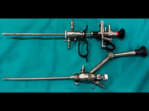
A surgical case using the MiLEP instrument set devised by Dr Felipe Figueiredo. The description identifies dissection and morcellation as the two parts of the case used to demonstrate the instruments. The accompanying commentary offers the surgeon's impressions of the equipment without reporting a comparative trial.
Documented details and sources
Only details explicitly stated in the sources. Missing information does not mean it did not occur.
- MiLEP
Real Life HoLEP cases: A MiLEP Case
Source checked: 2026-10-06.
Available transcript · English
Automatic YouTube captions, not clinically reviewed. Names, technical terms and numbers may contain errors.
Selected spellings of enucleation, morcellation and holmium terms have been edited. This is not a full audio or clinical review; the original captions remain the source.
They may contain transcription or translation errors; check the explanation in the video.
- Hi, this is Fernando Sancha. After a while, I wanted to show you because there's a lot of interest worldwide a millip case. There's a German company that has produced this 22 French um endoscope as well as the nephoscope with very interesting ideas uh improving the flow in small spaces and of course reducing the caliber uh in collaboration with Dr.
- Filipe figedo from Brazil and it is generating a lot of interest and I wanted to to show you. I spoke to the company they loan me some equipment to try it. I don't like the trigger you know that I use my fiber with the fingers and I think the trigger makes things a little bit more difficult in my opinion. although many people are used to it. But I'll show you this this case.
- This man had a catheter. He had a lot of bladder edema. He was on retention and he had a midsized prostate that I chose for for this opportunity. And as always, we're going to be able to see this uh milip equipment in action, but also uh of course we can refresh some of the tips and tricks that I usually use. So typically you know when I want to do the white line I mark the limit of the sphincter and you can find it easily on the sides probably 9 10:00 uh on the on the right side of the patient and probably 3 to 2:00 I do my first mark there and then I take that as a reference to go up towards 12 and to go down towards uh 6:00 keeping in mind that often the sphincter is a little bit tilted uh tilted anteriorly. So the anterior
- part of the sphincter is a little bit more proximal than the posterior part. Also as we know the sphincter has a horseshoe shape that uh makes it more important to preserve anteriorly than posteriorly. But here we are. This is the entry and as you can see I'm using the magneetto system with virtual basket at 2 JW 50 Hz which is my usual setting and I have uh the magneetto setting just for coagulation. Again here I'm going to the floor of the verontanum to look for the entry to the interface to the plane between the anoma and the capsule.
- I don't give it much thought at this stage. I just go in and try to find a depth that looks satisfactory, but I don't lose time trying to find out, you know, where is this mysterious beautiful plane. As you know, we cannot see this beautiful plane so much when we use this this new lasers because they provide very good first pass heasis.
- But that's the most uh beautiful I think you can see uh with these lasers you you get to see uh sometimes signs that you are in a in a nice interface between adoma and capsule there. I'm cutting the frenum of the montanum to connect both lateral spaces that I have open and I'm starting now to develop the posterior line as I usually do. Remember I think of the prostate like a little box. So I talk about the posterior plane, the lateral plane in both sides and the anterior plane. As you can see, I always put my scope aligned with the line of dissection. So in this case, I want to have the line of dissection in the middle of the screen to be able to move uh from side to side uh and develop this posterior plane. So
- here you can see that so far so good. We are seeing that uh there's a nice detachment of the plane as we progress from the verontanum towards the bladder neck posteriorly. The endoscope feels very light. Uh it is nice and I think the endoscope the the nucleiocope or the laser systocope that uh Felipe and this company have produced. I think it's it's uh wonderful in the sense that with a very small caliber you can do a normal job. Filipe is saying that he can do any size of prostates. I probably probably it's true. You you have to realize that with these new lasers the quality of of the first passis is very good. So the demands for irrigation I think are less and also if you do an in block technique we are irrigating you know a
- very small space and you don't need um a huge amount of uh irrigation power to to [Music] to be able to exchange the the amount of water contained in this very little space that we are forming between the and the capsule.
- So really I think the irrigation and the the view and the manuverability of of this scope is excellent. So I I have to say I like it and I think it is truly interesting how you can uh use it for cases where you know those cases where the urethra is smaller. I think this is opening new possibilities of being less invasive in in these patients. we have we have going we're going to publish uh a review of of retrospective review of 750 patients or something and I have to say that our rate of strictures using the 24.5 and 26 French uh endoscopes uh from Richard Wolf are extremely low. I I have to say also that I'm very very careful using the otus uh for the nvicular urethral um difficulties when I have to enter and
- I find the slightest you know tension at the miatus I would do a very gentle um throtomy and I have to say that we see very very rare uh rarely we see um strictures at the mat afterwards and then when I have the difficulty entering near the sphincter I try to use the a progressive dilator plastic uh progressive dilator that helps me enter very very gently.
- I'm always extremely gentle during the enucleation and during the introduction of the endoscope. But of course there's always cases with tiny urethraas urethra lumens and I think the 22 has come to occupy a space that was not occupied by any other laser before. This is a 12:00 fibers.
- You saw that I ascended from 6 to 9 sorry from 6 to 12 in both sides. And now we are connecting anteriorly. Trying to remove all the anterior tissue you know that we can find. So I don't try to leave uh tissue at 12:00. I try to be as aggressive as possible.
- But of course, you have to understand that before or many people who do an encircling technique to go up to 12:00 are going to need to be careful to see where they cut and how much tissue they leave because typically they have produced this uh mucosal flap at 12:00 and uh of course the flap is much more distal than our limit or the limit I mark at of the sphincter at 12:00. So this is an interesting thought. And here you can see that I am now developing the anterior line. So we had a posterior line. We developed both lateral lines a little to be able to mobilize the apex and to come up towards um 12:00 and connect and complete the early a liberation. And now I'm doing the anterior line detachment or this
- dissection. It's important if you want to reach the bladder neck, you have to put your scope on top of the adnoma and bring the adnoma downwards a little bit. And in order to have the mobility of the adinoma to do that, you have to cut this lateral attachments. You see this is the lateral lateral fixation that is going to uh fix the prostate and make the dissection of the anterior plane very difficult. So here I'm checking that the lateral plane and the lateral fixation at uh I don't know 10 11:00 is is being released cut so we can um mobilize better the adrenma. Sometimes I get a little bit distracted in the posterior area. You see I'm seeing this yellow stuff. So I'm realizing that maybe there is more
- admatus tissue than I thought. Maybe the plane I followed was not deep enough. So I am never shy to explore below the yellow tissue. And usually I find that I can remove more more admatus tissue. Often these are nodules, you know, adnotus nodules that grow into the peripheral zone. And you see whenever I see yellow I I try to explore and see if I can remove some more tissue. So I got a little bit distracted here. But the objective uh was to uh continue anteriorly. So let's see if I get back on track here. Coming up again trying to release this lateral attachments. Uh we call them hammock attachments also because when you put your endoscope at 12:00 you see they act like hammock attachments and they don't let you lower
- the adenoma with your endoscope. Of course I never do force. This is the other attachment. You see lateral fixation on this side. And now when we cut this lateral fixation when we put the adnoma on top of the sorry the endoscope on top of the the enoma uh we can access the bladder neck much more easily. So there you see don't try to go all the way to the bladder neck anterly without having cut this lateral fixation. You see now the the the prostate gets progressively more mobile and you can push it a little bit downwards and that will allow you to enter. In this case, this instrument in particular had a very tough very stiff uh trigger. So even when I when I wanted to use it, um it was a little bit difficult to push
- it in. It was like fixed and I had to do a harder push to the trigger and so it was not uh properly how do you say lubricated. Huh? Maybe. So maybe it needs to be used more often to to soften or smoothen uh that that movement.
- But now we have entered the bladder and that's a bladder neck. And you see I find much easier to use the fiber with my fingers. I have a lot of sensitivity. I can push it the fiber in and out. And this will help me find the working distance that I want in order to um you know do my dissection and find the effect I want to find at different stages in the operation. Here for example when I get closer to the bladder neck typically I want to be closer to the tissue because here what we want really is to perforate and um so many times you can do a little bit of touching you see and and cut the mucosa and open the bladder neck and you see that the the trigger is jumping a little bit very difficult to control so not not
- ideal and here in the posterior here area. I started questioning myself, is this a nodular prostate? Am I finding here that I found a plane that looked good, but could I be going deeper? And I'm never shy to to to to explore this possibility because often very often I find that uh we can find a deeper plane that can be can help you remove uh a lot of extra admatous tissue. I don't know if sometimes you know by exploring um you go deeper than you know the the the proper plane but in my experience I am usually more happy to explore and to question my depth of dissection trying to go deeper when I find yellow stuff that looks anomatous then you know regret it. So I usually explore and try to see
- how far I go trying to be aggressive uh removing all the anomatus tissue. If we leave anoma of course the anoma grows. It's a benign tumor. So we want to try to to be as aggressive as possible removing this anoma to prevent it from growing in the future and causing trouble.
- Uh some people say it's not important because the cavitation is still very good but I mind I don't know I want to be and also I have a lot of more fun if I am looking for these deeper planes very carefully using the energy very softly and carefully. Now, I had to say that this MIP instrument uh allows you to operate quite nicely.
- It feels very light and I I'm I'm going to wait to to to get one uh when I can have a freestyle, you know, port because I I hate the trigger. So, I know that some people are very used to it and they defend it. To me, the main problem of the trigger is that when you give a trigger to someone who has already done TRP, he's going to try to use it like a rectocope. So, he's going to be rotating. He he will have the fiber at six when he's at six in the adnoma. He will have the fiber at three when he's at three in the adnoma. He will have the fiber at 12 when he's at 12 and so on.
- So, and you see that I use the fiber very statically at 12:00 for most of the operation. I also work sometimes at 6:00, but I what I don't like is I don't like to rotate while I dissect because I think that movement is much more complex than what I'm doing now. You see, when I follow a line, it's very very precise.
- It's a very precise movement and that is going to help me I think much better to be precise and to be able of course of exploring these deeper planes you know playing with this these ideas and safely you know and the movement is more pure more simple and also if you look at my hands during the operation you will see that they're very static, you know, I just move around uh the fossa following these lines that I draw all the time around the anenoma, but I am not rotating the endoscope. I don't have to hold the camera so it doesn't rotate. So, it's much more simple manipulation strategy than the trigger or what the trigger makes you do. Of course, you can use the trigger like this. I mean not not
- um not rotating but uh the problem is that I don't have much freedom uh because usually I can push the fiber in and out very subtly to get the tissue effect that I want. So this is posterior and the case is nearly finished but I am still let's say trying to find out you see what is this? This looks like a nodule. That's a lot of anoma left. Maybe even there is some a little bit closer. That's a nodule. You see it's it's digging a little bit deep in the capsule. And I want to explore that depth to see if I can remove the whole nodule and not leave it there cut and attached to the to the capsule of the prostate. So there I am.
- That looks like yellow tissue. So I'm trying to find you see where is this yellow stuff? Yellow tissue admatus tissue and I'm trying to connect because of course you don't want to do many many cuts on the on the prostatic capsule and have like you know four parallel lines and then you don't know which line is which and where do you have to go. So I want always I want to generate lines that I can recognize later that I can connect with other lines you know and try to work in an in an orderly manner know and uh carefully here you see we're getting a little bit deeper in the in the capsule. It looks thin but it looks it looks okay. So I think that's that's the the nodular bit that we were finding. Now of course we have to
- connect the bladder neck come downwards towards the towards the midline trying to be as thorough as possible. Removing the tissue, trying to connect the lines. Let's see where we are. This can be a little bit confusing sometimes, but you see, I see this yellow tissue, and I think maybe I should go deeper. Try to remove that, too. You see, that's the idea. This is probably BPH tissue. There's more nodular stuff there. So, I'm going to come lower here. I have no reach if I keep the 500 clock. I'm going to use some of the 6:00 position to try to raise uh the this nodule up a little bit. Some people uh try to remove these nodules mostly at the end. So once they have finished the enucleation, they then
- go back to check if there is uh remaining nodules. I try to to find out you know if that's a correct correct depth when I am there let's say you know if I realize that there's yellow tissue I'll go to to pick it up and try to you know explore a little bit uh more deeply that depth you see sometimes it's you see that you're getting deeper in the capsule and sometimes you see signs of uh thinning of the capsule but it's typical that when there's a nodule growing against the peripheral zone, the peripheral zone will get thinner. So, here I am. I think I was starting to feel very happy that I did that because you see that there is this yellow nodule on top of me. Uh there you are. You see that nodule? So, this is
- what we are digging uh and extracting. And there we are trying to connect the lines. Uh you see that there is a thinner uh plane below. But of course this thinner plane is strong enough. Remember that we see a very very thin I mean the the the everything we see on the on the camera is highly amplified. The fiber measures half a millimeter. So you can tell that our field of view is probably four or five mm only. So whenever we see a nick in the capsule or a little hole or something, it's usually very small.
- And but of course I try to keep my energy close to the side of the anenoma in order to be able to dissect more. But um but um that was my wife entering the room. Sorry. And um you see I try to keep the energy up so I can uh ensure that I cut the fibers that join the adnoma with the capsule and [Music] um protect the capsule. You see that nodule that we have removed and now I want to connect the lateral aspect. You see if I see that in some areas the capsule is a little bit deep. I will fire a little bit closer to the anoma to correct my my targeting and to avoid following the deep plane into a deeper plane. There we are. So in principle for this first part of the operation, I think this instrument is very very usable. I'm not
- sure how would I feel. I don't have as much experience with this instrument as Filipe has, but I'm not sure how would I feel doing a very very large gland probably. And what I'm thinking about this business of miniaturaturization of the scopes, we have seen the same with uh stones, you know, we went from perccutaneous surgery with standard sizes to mini perk and ultra mini perk and people were enthusiastic about it uh because it would be less aggressive for the patient. it would cause less trouble with the entry into the kidney and so on. But then I think there has been a let's say return to to to to to be sensible. No, there are cases where mini perk is wonderful.
- There are cases where people still use the standard um instruments because the size of the stone is too too big you know and then you don't need to suffer so much yourself uh trying to do a very difficult job through a very small instrument. So I think millet is here to stay. I think most of us surgeons who do a lot of holyps will want to have a smaller uh sized instrument.
- And I also think [Music] that we will probably still choose um a little bit bigger scopes for very very difficult cases. I don't know. This is my this is my prediction of of what is going to happen.
- So kudos to to Figuredo and kudos to the company who did this this endoscope. I think the endoscope is very good and as soon as we finish our enucleation we will be able to move on to more solution and that's a little different story and you will see but in principle using this scope is very nice. It's a little bit delicate.
- It's small. uh if you use the smaller size 18.5 French I think this is a much more delicate instrument you cannot use um very high power because um even the tip I mean could could be damaged by having this effect of the energy so close to it and now this is the hemoasis and trimming phase if I find tissue that looks adomatus of course I will want [Music] to take it out. I want to make the capsular fossa as perfect as possible. And as I said, I didn't find any problem operating with this uh little instrument except what uh you know my preference regarding the not using the the trigger. I think the trigger it's not so comfortable and if you are very comfortable with the trigger you should try to use your
- fingers because it's a it's a new feeling and after some cases of feeling you know a little strange without your trigger. uh you might find that uh the freedom you have is amazing and the sensation you know you hold the fiber with your fingers and you have a lot of control and also it allows you to do this rotationless dissection you know keeping the fiber at 12:00 uh during most of the operation. So here we have you can see the mucosa of the sphincter is not suffering it's not suffering from the um cheese grater effect or it's not suffering from the the size you know causing a little bit more eskemia. So all these factors I think are extremely reasonable and uh also I find that uh the argument that
- Felipe gives to his patients is very convincing. you know he shows him them three sizes of instruments 18.5 22 and uh 24 26 and he says which one would you like me to use in inside your penis you know and then the patients are horrified and of course they want the smallest size no so that's an irrefutable argument no we all would want uh to have a thinner endoscope in our urethra but as I said we reviewed 750 patients and the rate of of stricture in our hands was very very very small. So I think there's no how do you say and also there's no way to know if we will get less strictures when we use a smaller size endoscope. It is logical to think that it is going to be difficult to prove it scientifically in
- a study because if we have something that happens with a relatively low incidence uh we will need to use huge numbers of patients to prove you know that there is a clear advantage uh of using smaller sized endoscopes. So I think the enucleation is finished and the morflation is going to take place.
- Now this is a change of instruments. I am using the I'm changing the laser systocope for the morsoscope. But this Morscope in order to have an overall small size of of the sheath and uh in order to to have good irrigation.
- This Morscope has a problem and that is that it uses a fiber optic lens. So this is like traveling to the past you know when we did irroscopy with these kind of camera systems. So I think probably with this instrument moreation is going to be a little bit more tricky. There's people around using this instrument saying that they let's say suffer or struggle a little bit more but they manage to do more but uh I think the the reduced um irrigation uh Philipe says that if you use two inflows connecting you know four bags with two lines uh as the inflow if you use uh two inflows you will get a similar irrigation as using a 24.5 from Richard Wolf uh using one inflow which is what I usually use but then there is this quality of
- the image uh problem I have heard from some surgeons who are using this instruments that have let's say stopped morcellating with this device and when they have to morcellate they upgrade to 24.5 5 for morcellation.
- I'm going to give you a clip with the normal or maybe I could insert it here. Just a moment. So here I inserted a clip of a morcellation with a 24.5 uh endoscope and you can see that the the quality of the image is a little bit uh better uh using a proper road lens than using the fiber optic lens. So here I think this is the weakest um weakest uh morellulation. I think if you don't have absolutely excellent hemostasis you will probably not see so well because of the lack of of visibility. When I have trouble with visibility, I normally I use one uh inflow in my morscope and it's enough to keep it simple and uh finalize the morcellation. But um I have to say that you can always insert a second irrigant, you know, to
- to to get even better inflow when things get a little bit difficult. But overall, and I think now that the video is is uh nearly finishing, I think that it is a step forward. It is a nice it's a welcome option. As I said, I'm going to wait until the company produces a freestyle element. I'm sure that all the companies would get will get interested in in miniaturaturizing the instruments for HOL. as HOL is becoming more and more popular in the world because you know that up to now it has been quite a niche operation and that's why we have been abandoned by companies who have not developed uh new instrument for us also they tell me that developing new instruments is a pain as the regulations and the you know
- C mark and FDA approval are a real pain and difficult. So, for the moment, I'm going to stick with my 24.5 and 26. As I said, um I wish I could have an instrument like this without a trigger because the trigger makes it really unpleasant for me to to work. But uh I think it's a great u advancement and we have to congratulate Figuredo and the company the German company that has produced this instruments uh that was kind enough to loan the equipment for me to try and here well this is a video and this is my opinion and these are my ideas. So, I hope you enjoyed the case and see you around. All the best. Take care. Bye-bye.