
Surgery
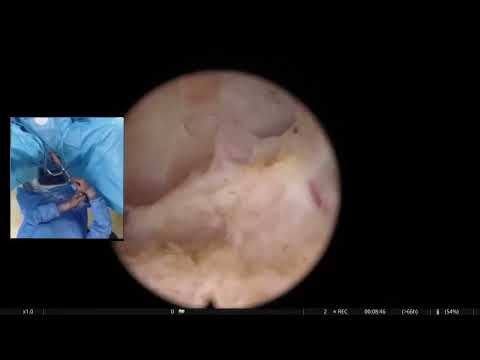
RL HoLEP: a patient with a right complete duplex system draining in the prostatic urethra
Dr. Fernando Gómez Sancha · ICUA
Dr. Fernando Gómez Sancha · YouTube
Language: English. YouTube audio metadata.
Surgical content for professionals. Contains footage of an operation.
Playback connects to YouTube. Your search query is not sent to the player.
About this video
An en-bloc HoLEP with an unexpected ectopic ureter opening into the prostatic urethra. The title identifies a complete right-sided duplex system, while the description explains that the intraoperative approach was adapted to preserve the ectopic unit. The teaching focus is recognition of an unexpected ureteral orifice and the resulting operative decisions.
Documented details and sources
Only details explicitly stated in the sources. Missing information does not mean it did not occur.
- En bloc
In this video, I present a rare intraoperative finding during a HoLEP en bloc procedure: an ectopic ureter draining into the prostatic urethra — a congenital anomaly that is exceptionally uncommon in adult patients.
- Ureteral duplication
RL HoLEP: a patient with a right complete duplex system draining in the prostatic urethra
- Ectopic ureter
Intraoperative Discovery of an Ectopic Ureter Draining into the Prostatic Urethra During HoLEP
Source checked: 2026-10-06.
Available transcript · English
Automatic YouTube captions, not clinically reviewed. Names, technical terms and numbers may contain errors. Check them against the video; complete audio coverage has not been verified.
They may contain transcription or translation errors; check the explanation in the video.
- You can stay everybody has a 14 not everybody knows there's no circumcision attit
- It's a little bit tight. The fiber
- make a small thermal ring. this mucosal vein is very common. So I'll try to minimize the amount of uh damage put by the detection and then position maybe that's enough to be able to dilate it very carefully. Also, you can use the fiber as a guide wire to help you in there. Sometimes when you have a tight, you can use the fiber like wire. Okay, there we are. This is a 16. This is a false passage or some interesting cavity there. Maybe
- could be a unit maybe a topic here and there. Let's see. Looks like an ectopic here. So, it's a very nice case. It's a very interesting case. So, we come out. You see the edge of the center here and maybe it's difficult to tell maybe around there. It's usually symmetric or slowly symmetric when it's
- uh When you have uh one let's say congenital formation, you can have more. Sometimes we see that we enter the plane and there's very strange looking tissue is usually malarian remnants on the it's not very common. Just see a couple of pieces in my life. Okay. So here again we enter
- Good. Here we are.
- Try to connect the posterior line of dissection.
- It's a little bit deep there. Maybe trying to go deep down there. Let's go to aexics. again the white line so we get more information and more axis. Then we come here try to start the lateral plane. Here
- I'm noticing some resistance. So when I go back you see we find that there is some touchment here. We remain above the ab the normal below the spin curve. Move out there as much as we can. Double look here. Now we're going to come from here. Time to go up up once. Sometimes the line that we cut remains below. You see we cut from here. And now going up there to try to find the uppermost plate. It's not 12:00 fibers. That's okay. We release that
- 12:00 fiber. Sometimes we get a little bit better mobility. So that's why I cross over a little bit to the other side to get some some more mobilization. There we are.
- Okay. Okay. So this side looks quite good here, here and here. It's also 12:00 here. Now we come out and check where is the white line here. See the edge white line. Let's deepen a little bit center here. This is the lateral natural wing. Let's see.
- And now here again we are coming towards the look. This is all white. The fact that it's white tells me that we've been here before and also it's a safe safe place. It helps a lot with orientation because we don't need to go back look at this paper that come out again you know Just
- we are coming around trying to define the L line in the interior. here. It's looking good. And now we will check what happens at 12:00. Come out. See, we have this 12:00 fers. We'll try to stay at leader. the connection at 12. This is now the antior line that connects to the lateral line here. And as we come up up
- this rejection now you're running connecting to the other side. Let's see what we have here. Here we went a little bit further. Stay up and come and see. Sometimes when we are antidiorly here, we want to reach the the bladder neck prostate doesn't descend very well because there's lot of fixation. So we come now to the sides. Try to release the lateral fixation here. These are things that uh we didn't know before. You know what I mean? When we
- started doing these are things that you realize no over time. It's not so easy to you come to the conclusion why am I am I not getting access to the to the neck. You see now you see how we can bring it down more because we release these attachments. We call them the hammock attachments because they're like holding prostate very very rigidly in place. There's no way to to move it to to change the so I don't know what kind of evaluation this man had of the upper tract but
- we have to see because probably he has a funny let's say double system or something also I didn't pay attention to the to the U inside the bladder we need need to Now when we enter we need to check to see if there's one you know normally positioned urator and then another ectopic urator only the upper y no it's dilated [snorts] and uh it's coming to the mix rather than the on. It's uh leading to the rea. There we go.
- Now that we see we were able to descend a little bit the enorma to get optimal entry.
- It's going to look So this one it's okay. There's another one there. probably we have a third urer coming to heration. So yeah system I think with one of the normally the upper mo let's say comes the urer is going to to to be positioned lower but in this case it's intra prostatic and intra we should see it when we do sticker plane I think let's see because we saw that there was like a tunnel going This is normally the unit there, you know, the dumping unit there.
- [snorts] Um, Alex, can we call see if we can bring home
- Because maybe it's not dated. Maybe you don't realize that yes system. No, you don't notice something that you need. You have to suspect. No, that There's a second uh normally you can see the two separate uh fat component in the ultrasound image. Now when you see the be the kidney normally uh yeah this man has a very funny very strange cold here. This is prostate. this hole and this looks like an ectopic urer coming up here. See looks like a urinating in the prostate. So I think it has a duplex system the upper part of the kidney draining here. Yeah.
- So we're going to do the posterior plane now. We probably cut into the urer and the ur will remain in the proic fossa. But it would be nice to do an ultrasound of this man lucky to see if we can see more and understand better. In any case, I think with this interoperative findings, he should have a CT scan maybe with with contrast. Okay. Just to know there's nothing here. Do you think it's possible to bring uh here? Okay. When we finish. Yeah. Yeah. Yeah. I will tell This is posterior plane. PL can see here. We're trying now to come up
- here. You see this? This is very interesting. Now, when you dissect in the midline posteriorly, you see you come to this point and it looks like the plane wants to go up up here. You see? Looks like it wants to go there. And then when you do the lateral, you can see that there's much more tissue here. You see that? This is this this tissue here is what I call the ligament because basically this is the plane of developed by the middle when it grows uh pushing downwards. You know what I mean? This is the middle plane below the middle. No. But then when you come to the side, you realize there's much more lateral because the lateral lo has grown and pushes outwards you know. So in between we have sometimes this
- avalent discrepancy and this is what I call the ligament. I cut the ligament. Okay. You see you see that kind of that kind of uh discrepancy in the plane. Believe that you can believe that there is more tissue naturally. You know it's not that you have two wrong claims or something like that or one claim is right and the other one is wrong. It is because the prostate is not a proper billard at all. You know it's not absolutely hysterical. It's uh sometimes irregular sometimes you get theoma. It has like three loes and this is the shape of the lateral lo coming down here. But then of course
- this is something we find quite often. Here we are a little bit deep. Let's see. Looks very yellow the tissue. That's why I'm getting a little deeper. I think it's adding on. I would like to take it out. But let's see. This side we have done a lot in this side. Let's go to the other side now and do a little bit more on the others. We line stay up with our fiber.
- See how this connects now to the lateral tissue here.
- Let's try to come from the sides. I think if we do the sides very well when we reach this middle part here I think you will be seeing a little bit better what's going on with this uh there we are going down this side trying to release the lateral aspect here B on the line. The line of dissection.
- It's jumping a little bit. And now we're entering the urator. You see this is the urer dilated urer. You see that goes up to the kidney. Now so maybe we should lower the backs a little bit. Maybe release the pressure. Yes. You bring the bags down. Backs down. Back down. You only go so much pressure. It's of course looks like uh the left sorry right right side
- to focus sorry target the beam against the noma. Try not to go deeper on the capsule. Try to mobilize a little bit more towards the other side so we can see a little bit better what's happening.
- looks a little bit deep but still acceptable. Sorry, it's jumping. Jumps. There's this attachments here. Can I do this?
- Every millimeter we release gives us more mobility and more access. It's a process. We need to be careful and try to gain access and gain mobility trying to stay inside if you can. So pointing the edge of the other because if you point down here, of course, we're going to deepen into the capsule much more. Now, this side is looking good. I think really getting towards the bladder there. Let's see
- really well here. Now need to connect this. We have opened the entry node to the ureric drainage points.
- What's up here? See, so now we can drain into the capsule rather than into the procedure. We need to be careful that the catheter doesn't go up here. Huh. When we put it, so I'll try to keep the point um towards the left side at least the patient. less possible to put it into the military opening. So very interesting case.
- So hopefully it's uh you is okay here. The other should be also okay around here here. And then the third one is here. No. Try to do some more. Nice. Very beautiful case.
- come out. You should see the sphincter. You see the muzza is a little bit skinny by the movements, but it's covering the the sphincter everywhere. So, it's looking good. But I think will not be an issue from this one. Wonder how much urine this kidney upper moment is is producing. Maybe not so much. That's a little bit deep here. But we're going to try to finish early now. I don't think it's horrible. It's just a little bit of deepening there opening and Let's see because often the bleeding comes in the post.
- We're keeping the bags low so we don't have a lot of pressure going out to the kidney. Let's see how
- Obviously
- vessels are important for visibility during feeling that there something's going on here. the moment setting. So we very it's very reliable you know when you usually does it very well here again you see you don't spend the time to do the then you jump into more early and then you have trouble
- I think it's getting a bit dark.
- This is the deep area the flow. Where is it? Here. Okay, this is like this might be a bit of a localization when in Madrid we have an ultrasound transctal ultrasound in the operating room. So if you stick the ultrasound probe in the rectum you can see there isation sometimes there's a little where you are you don't want it to be a lot but when you're working with no pressure
- and you're going to finish fast so as I said before many experts do not care too much about preparations because of course they are fast and the chances that these preparations are going to cause massive traational water are go. But then when you're learning, if you do a profession, you have to be careful because it might take you a long time to to get to the end of the procedure and there might be more time, you know, to build up more expensation. So you need to be careful when you evaluate, you know, how important it is to have a preparation
- bubbles as them. There we are again. Ability is not so good because we have very little pressure. We need to be careful. Uh blue wall elevated by by
- Yes, it's possible to plug it and turn it on.
- Okay, is going well. I'm very static in a safety position. We elevated the pressure again to have some more pressure gulation. Hopefully it's not a problem.
- Final two seconds here. This looks like piece or this is capsule behind the capsule. Let's uh put a path there. The upper moatic I think of the kidney. Check is this is going this way or this way. So normally when when you do ultrasound I'm doing it with the wrong hand. Sorry. You you can see that there's like two differentiated fat parts. You see this one in the in the upper part here. You see that it's a little bit dilated probably after the dilotation that we
- have caused. And then there's another lower uh you see. So probably he has a duplex system. No effect.
Available transcript · Spanish
Subtítulos en español publicados por el canal. Pueden ser una traducción asistida por IA; no se utilizan para inferir atributos clínicos.
They may contain transcription or translation errors; check the explanation in the video.
- Vale.... está un poco apretado dame la fibra, ya sabes, puedes ver cómo aparece. esto es un pequeño anillo mucoso. Estos anillos mucosos son muy comunes. Intentaré minimizar el daño causado por la disección Y luego la incisión, quizá eso sea suficiente para poder dilatarlo con mucho cuidado También puedes usar la fibra como guía para ayudarte a entrar A veces cuando está apretado, puedes usar la fibra como una pequeña guía.
- Vale. Ahí estamos. Esto es un 16-ohm. Esto es un falso paso o alguna cavidad interesante ahí. Quizá un utrículo Quizá podría ser un uréter Quizá un uréter ectópico Quizá un uréter ectópico Vamos a ver. Parece un uréter ectópico, así que es un caso muy bonito. Es un caso muy interesante. Salimos. Puedes ver el borde del esfínter aquí Vale. Y quizá es difícil de distinguir. Quizá por ahí. Suele ser simétrico
- A veces cuando tienes una, digamos, malformación congénita, puedes tener más. A veces vemos que entramos en el plano. Y hay un tejido de aspecto muy extraño. Suelen ser restos müllerianos en la próstata No es muy común. Solo vemos un par de casos en nuestra vida. Vale. Así que aquí de nuevo, entramos. Aquí estamos Intentar conectar la línea posterior y la disección.
- Está un poco profundo ahí quizás. Intentando ir profundo ahí abajo. Vamos al ápex. De nuevo, la profundización. Profundizar un poco la línea blanca para obtener más información y más acceso. Luego venimos aquí. Intentar iniciar el plano lateral. Aquí estoy notando algo de resistencia. Cuando vuelvo atrás, vemos que hay alguna inserción.
- Aquí nos mantenemos por encima del adenoma por debajo del centro. Así que subimos ahí todo lo que podamos. Esto son las 12 Aquí. Ahora vamos a venir desde aquí. Hora de subir subir subir A veces la línea que hicimos para cortar queda por debajo. Ves que cortamos desde aquí. Y ahora voy a subir para intentar encontrar el plano más alto. Esto son ahora las doce Fibras de las doce Eso está bien. Cuando liberamos las fibras de las doce A veces conseguimos un poco más de movilidad. Por eso cruzo y conecto al otro lado para conseguir más movilización
- Aquí estamos Aquí estamos Vale Así que este lado se ve bastante bien Aquí estamos anterior También son las doce aquí Ahora salimos y comprobamos dónde está la línea blanca. Aquí. ¿Ves? Borde del esfínter Línea blanca. Se ha profundizado un poco. Eso está ahí dentro. Aquí. Este es el plano lateral
- Vamos a ver Y ahora aquí de nuevo venimos hacia las doce Esto está todo blanco. El hecho de que esté blanco me dice que ya hemos estado aquí antes, ¿no? Así que es un lugar seguro Ayuda mucho con la orientación porque no necesitamos volver y mirar el esfínter salimos, De nuevo, ya sabes, estamos rodeando
- Intentando definir la línea lateral y empezar la anterior Se ve bien. Y ahora vamos a comprobar qué pasa a las doce Salimos. Aquí tienes estas fibras de las doce Tiene libertad. Intentaremos quedarnos anterior. Así que esta es la conexión a las 12 Esta es ahora la línea anterior que conecta con la línea lateral aquí. Y vamos a ver... subir, subir, subir. Tenemos esta conexión ahora, ¿no? Aquí en la línea anterior. Está conectando al otro lado.
- Vamos a ver qué tenemos aquí. Aquí fuimos un poco más lejos Quedarse arriba. Y capsular. Vamos a ver. A veces cuando estamos anteriormente aquí y queremos llegar al cuello vesical, La próstata no desciende muy bien porque hay fijación lateral Entonces venimos al lado para intentar liberar la fijación lateral aquí. Estas son cosas que no sabíamos antes. ¿Sabes lo que quiero decir? Cuando empezamos a hacer... Estas son cosas que te das cuenta con el tiempo. No es tan fácil de... Llegas a la conclusión, ¿por qué no consigo acceso al cuello vesical? ¿Ves? Ahora ves cómo podemos bajarlo más porque liberamos estas inserciones.
- Las llamamos las inserciones en hamaca. Porque están como sujetando la próstata muy, muy rígidamente en su sitio. No hay forma de moverla, de cambiar la... Así que no sé qué tipo de evaluación tuvo este hombre del tracto superior. Pero tendremos que ver, ¿sabes? Porque probablemente tiene un, digamos, sistema dúplex o algo así. Tampoco presté atención al orificio ureteral dentro de la vejiga Necesitamos comprobarlo. Ahora cuando entremos, necesitamos comprobar. Ver si hay uno, ya sabes, uréter normalmente posicionado Y luego otro uréter ectópico Probablemente la porción superior ¿No?
- está dilatado Y viene hacia la uretra en vez del trígono Drena hacia la uretra. Drena hacia la uretra Ahí vamos Ahora que entramos en la vejiga, vemos que pudimos descender un poco. El adenoma para conseguir una entrada óptima. Va a verse... Así que este está bien. Hay otro ahí. Así que probablemente tenemos un tercer uréter que llega a la uretra
- ¿cómo dices? Es un sistema dúplex, creo. Es uno de los... Así que normalmente la porción superior, digamos, viene... el orificio ureteral va a estar posicionado más abajo Y en este caso, es un uréter intraprostático Deberíamos verlo cuando hagamos el plano posterior, creo Vamos a ver. Porque vimos que había como un túnel yendo hacia arriba. Que probablemente es el uréter Ya sabes, el uréter ectópico Alex, ¿podemos llamar al Dr. Botsevski? A ver si podemos traer un ecógrafo para comprobarlo. ¿Vino? No, por favor. Porque quizá no está dilatado.
- Quizá no te das cuenta de que tiene un sistema dúplex. Haces una ecografía en la consulta Tienes que sospechar que hay un segundo... Normalmente puedes ver los dos componentes grasos separados en la imagen ecográfica Cuando ves la judía El riñón con forma de judía La judía del riñón. Normalmente. Alex, Sí. Este hombre tiene un agujero muy curioso, muy extraño aquí. Esto es próstata. Esto es agujero. Y esto parece un uréter ectópico subiendo por aquí ¿Ves? Parece un uréter drenando en la próstata. Así que creo que tiene un sistema dúplex. La parte superior del riñón drenando aquí. Sí. Así que vamos a hacer el plano posterior ahora. Probablemente cortaremos el uréter y el uréter quedará en la fosa prostática
- Pero estaría bien hacerle una ecografía a este hombre. Para ver si podemos ver más y entender mejor. En cualquier caso, creo que con estos hallazgos intraoperatorios, debería tener un TAC. Quizá con contraste. Vale. Solo para saber. Sí. No hay nada que podamos hacer. Probablemente solo comprobar. Sí. ¿Crees que es posible traer un ecógrafo? ¿Aquí? Vale. ¿Cuando terminemos? Sí, sí. Sí. Yo aviso. Este es el plano posterior. Plano posterior. Puedes ver aquí. Voy a intentar ahora subir. Y aquí. Mira, esto es muy interesante ahora. Cuando disecas en la línea media, posteriormente, ves que llegas a este punto. Parece que el plano quiere ir hacia arriba aquí. ¿Ves?
- Parece que quiere ir ahí. Pero luego cuando haces el lateral, puedes ver que hay mucho más tejido aquí. ¿Ves eso? Esto, este tejido aquí es lo que yo llamo el ligamento. Porque básicamente, este es el plano del, desarrollado por el lóbulo medio cuando crece empujando hacia abajo. ¿Sabes lo que quiero decir? Este es el plano del lóbulo medio. Debajo del lóbulo medio. Pero luego cuando vienes al lado, te das cuenta de que hay mucho más lateral. Porque el lóbulo lateral ha crecido y empuja hacia fuera. ¿Sabes? Así que, entre medias, tenemos a veces esta discrepancia aparente. Y esto es lo que yo llamo el ligamento. Corto el ligamento. ¿Vale? ¿Ves? Ves ese tipo de, ese tipo de discrepancia en el plano. Créete eso. Puedes creer que hay más tejido lateralmente. ¿Sabes?
- No es que tengas dos planos equivocados o algo así. O un plano está bien y el otro está mal. Es porque la próstata no es una bola de billar perfecta ¿Sabes? No es absolutamente esférica. A veces es irregular. A veces sacas el adenoma y tiene como tres lóbulos. Y esta es la forma del lóbulo lateral bajando aquí. Pero luego por supuesto, y esto es algo que encontramos bastante a menudo. Aquí estamos un poco profundos. Vamos a ver. Es muy amarillo, este tejido.
- Por eso estoy profundizando un poco más. Creo que es adenoma, me gustaría sacarlo Vamos a ver Este es este lado, tenemos mucho en este lado, vamos al otro lado ahora Para hacer un poco más en el otro lado Nos gustaría quedarnos arriba con nuestra fibra Vale. Mira cómo esto conecta Ya sabes, con el tejido lateral aquí Vale.
- Vamos a intentar venir desde los lados Creo que si hacemos los lados muy bien, si llegamos a esta parte media aquí, creo veremos un poco mejor. veremos qué pasa con este otro uréter Ahí abajo. Aquí estamos Bajando, decidimos intentar liberar los aspectos laterales aquí tropiezo en la línea, la línea de disección... saltando un poco ahora estamos entrando en el uréter, ¿ves?
- este es el uréter....uréter dilatado... ¿ves eso? Sube hasta el riñón deberíamos bajar las bolsas un poco, quizá soltar la presión, Lubo, ¿bajas las bolsas? no quieres tanta presión Parece el izquierdo... perdón derecho, uréter del lado derecho Intentas enfocar, perdón, apuntar el haz contra el lado del adenoma
- Intentar no ir más profundo en la cápsula... empezar a movilizar un poco más hacia el otro lado para que podamos ver Un poco mejor qué está pasando un poco profundo pero aún aceptable
- perdón está saltando, salta porque hay estas inserciones aquí cada milímetro que liberamos nos da más movilidad y más acceso, es un proceso, deberías ser cuidadoso e intentar ganar acceso y ganar movilidad intentando quedarte dentro si puedes apuntando contra el borde del adenoma porque si apuntas aquí abajo por supuesto vamos a profundizar en la cápsula mucho más este lado se ve bien, creo que casi llegamos al lecho vesical, vamos a ver
- bueno aquí ahora necesitamos conectar esto, habíamos abierto la entrada al punto de drenaje ureteral Eso está aquí arriba Ahora está drenando en la cápsula en vez de en la uretra prostática, tenemos que tener cuidado de que el catéter no suba aquí, así que intentaré mantener la punta hacia el lado izquierdo al menos
- para que el catéter quede proximal a ese drenaje ureteral es un caso muy interesante así que esperemos que este orificio ureteral esté bien aquí, el otro debería estar también bien por aquí aquí y luego el tercero está aquí, vamos a hacer algo de hemostasia y morcelar
- Caso muy bonito, salimos, deberías ver el esfínter, ves que la mucosa está un poco isquémica, por los movimientos, pero está cubriendo bastante el esfínter, así que acerquémonos un poco aquí Creo que están por todas partes, así que se ve bien, pero creo que la continencia no será un problema para este hombre Me pregunto cuánta orina está produciendo esta porción superior del riñón. Quizá no mucha. Está un poco profundo aquí, pero vamos a intentar terminar pronto, y no creo que sea una perforación horrible. Es solo un poco de profundización ahí.... apertura ureteral...
- Vamos a ver la mucosa, porque a menudo el sangrado viene de la mucosa. Estamos manteniendo las bolsas bajas, para no poner mucha presión subiendo al riñón. Esperemos... Vamos a ver cómo... Obviamente la hemostasia es muy importante para buena visibilidad durante la morcelación
- sintiendo que algo está pasando. Aquí está el ajuste del Magneto Es muy fiable. Ya sabes, cuando coagulas normalmente coagula muy bien. Aquí de nuevo, ves. Si no dedicas tiempo a hacer hemostasia, entonces saltas a la morcelación muy pronto, y luego tienes problemas. Ahora creo que está mejorando. Esta es la zona profunda.
- Esto es el ureteral... no ¿Dónde está? Aquí, más lateral. Vale. Esto es así. Podría haber un poco de extravasación local. En Madrid, tenemos un ecógrafo, un ecógrafo transrectal en el quirófano, así que si introduces la sonda ecográfica en el recto, puedes ver si hay extravasación. A veces hay un poco. Ahí estás. No quieres que sea mucho. Pero soy rápido como operador, y voy a terminar rápido. Así que, como dije antes, muchos expertos no se preocupan demasiado por las perforaciones porque, por supuesto, son rápidos, y las probabilidades de que estas perforaciones causen una penetración masiva
- de agua son bajas. Pero cuando estás aprendiendo, si haces una perforación tienes que tener cuidado porque puede llevarte mucho tiempo llegar al final del procedimiento, y puede haber más tiempo para acumular más extravasación. Así que tienes que tener cuidado cuando evalúas la importancia de tener una perforación las burbujas normalmente puedes aspirarlas con el morcelador Aquí estamos de nuevo. La visibilidad no es tan buena porque tenemos muy poca presión. Tenemos que tener cuidado. Lubo, sube las bolsas... bolsas arriba
- ¿Trajeron el ecógrafo, sí? ¿Es posible enchufarlo y encenderlo? Vale. La morcelación va bien. Estamos estáticos en posición de seguridad. Subimos la presión de nuevo para tener más presión durante la morcelación
- Esperemos que no sea un problema. Ya sé. O esto parece un trozo o esto es cápsula. Estamos en la cápsula. Creo que hemos terminado Vamos a poner un catéter. La porción superior, creo, del riñón.
- Déjame comprobar. ¿Esto va en esta dirección o en esta? Normalmente cuando haces ecografía, porque lo estoy haciendo con la mano equivocada. Perdón. Puedes ver que hay como dos partes grasas diferenciadas. Ves esta en la parte superior aquí. ¿Ves eso? Está un poco dilatado, probablemente después de la dilatación que hemos causado. Y luego hay otra más abajo, ves, así que probablemente tiene un sistema dúplex. ¿Hmm? No, entonces, perfecto.