
Surgery
RL HoLEP case 36, presented at Urotech Congress in Istanbul, Turkey
Dr. Fernando Gómez Sancha · ICUA
Dr. Fernando Gómez Sancha · YouTube
Language: English. YouTube audio metadata.
Surgical content for professionals. Contains footage of an operation.
Playback connects to YouTube. Your search query is not sent to the player.
About this video
A HoLEP case prepared for the Urotech Congress in Istanbul, Turkey. The author explains that technical problems prevented the planned live commentary, so this upload includes the commentary afterwards. The written description does not identify particular case measurements or clinical outcomes.
Documented details and sources
Only details explicitly stated in the sources. Missing information does not mean it did not occur.
No clinical case details have been extracted for this video.
Original title and description on YouTubeSource checked: 2026-10-06.
Available transcript · English
Subtitles exported from the original-language YouTube Studio editor. Not clinically reviewed; source identity and timestamps verified, full audio coverage not verified.
Selected spellings of enucleation, morcellation and holmium terms have been edited. This is not a full audio or clinical review; the original captions remain the source.
They may contain transcription or translation errors; check the explanation in the video.
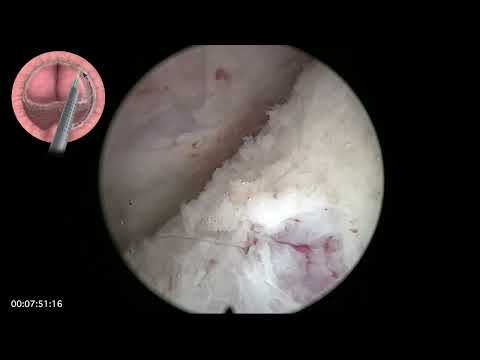
- hello this is fernando romer sanchez and this is a video i presented for the eurotech meeting i was supposed to connect via zoom and comment on it online but there was some technical problems and i was a little bit late to connect so i couldn't discuss this during the congress and i'm going to try to do that now so and i'll put this video in the channel so it is available for those of you who couldn't maybe understand everything or couldn't hear from me what what i intended to to explain this is a 62 year old man with acute retention of one liter and 50 50 cc prostates and this we're going to use the virtual basket laser to perform an unblocking nucleation i will try to reflect on the step-by-step approach
- the video lasts about 25 minutes i chose this case because this was the surgical time for this for this patient three lobe and so the initial step of the procedure is to mark the line the white line around the sphincter to to try to cut the mucosa so it doesn't break uh later on with manipulation so that the mucosa will remain on the sphincter so here of course you have to get close to the sphincter and try to you know use the anatomical landmarks to [Music] to perform this this incision i was in a congress that someone said this is a you know artificial line um how are you sure where are you doing this line and of course with experience you can get to identify you see the edge of the sphincter
- and make a line that is very very close to the real thing if if you can uh see very well the first thing we do is to [Music] enter the the the plane right at the floor of the montanum you see this is on the right side of the patient so you go to the floor there and you incise and you move progress laterally i like to cut this distal fibers if i can first because that will reduce the traction on the sphincter when we try to put the scope below the apex of of their norma there you are so that's the entry into the plane and then we're going to connect the planes by cutting the frenulum of the vera montanum and that's the cut so i will concentrate on on fabricating a line of dissection that goes from side to side and i'm going to aim with my fiber
- against the line so this is the the line from side to side and i like to keep my movements very very wide you know i don't like to work deepening in a very small area but just move you know from side to side to have a wide dissection effect and also because i want to protect this line of dissection as you will see in this case we don't get to see a very beautiful plane so i will have to judge by looking at the anatomy by looking at the characteristics of the plane to see if this is a good plane or not it looks maybe we're going a little bit deeper than the the proper plane but it is dissecting excellently well and i i don't really care too much about uh seeing the the beautiful plane that sometimes we can see but not in every part of
- the prostate and not in every patient here you see this is the line of the mucosa there was a small split starting to happen but i i will try to to correct that of course and this is the mobilization of the apex i'm trying to do a little bit of mobilization of the lateral aspects of the apex this is the face of mobilize and connect i call it mobilize and connect because you want to gain some access and there we're going to remain below the sphincter trying to deepen i'm going to cut a little bit on the adnoma you see this cut is going to give us access to the proper plane so initially we cut on the adenoma and then we go to look for the good plane the initial cut will give you access and we'll protect the sphincter on the mucosa and then of course
- we have to ascend in our dissection uh to try to remain eccentric so that we don't cut through the uh horizontally and leave anomalous tissue at the apex so here we're coming up upwards and i'm trying to release not only lateral uh lateral tissue but also a little bit of the anterior plane on the way to the bladder neck because if we don't mobilize the apex it's very difficult to reach the 12 o'clock area here again i'm cutting on the adenoma this is my first cut that first cut doesn't mean that i'm going to follow that plane i'm going to follow the correct plane but the first cut gives us very good access to this plane a little mobilize and connect phase here where i mobilize the posterior apex on the left side and
- connect the line because i want to fabricate a circumferential line that will help me navigate the procedure with a pretty good idea of the depth of of the nucleation if you look at an mri you will see that the line of separation between surgical capsule anama is quite quite uniform so here i'm doing another cut now more horizontal you see just below the sphincter trying to get better access and then i'm going to go to the look for the good plane and try to continue my dissection upwards slowly and progressively see that's coming up coming up coming up looking back at the apex i would like to cut the apical attachments first because what i don't want to do is to dissect inside come up and put some traction on this area so i'd rather cut
- the distal axis first and then when i have good access then i will progress with my dissection trying to [Music] get closer and closer to towards 12 o'clock so here here you see this is a 12 o'clock region this is the plane we had on the right side coming up and now i'm going to go from one side and the other and try to cut the 12 o'clock fibers here you can see 12 o'clock fibers this is a small nodule right at the apex anteriorly so we want to go above that and having dissected the anterior plane from both sides it is now possible to cross from one side cut the 12 o'clock attachment and reach the the plane on the other side so you will see how the previous dissection the previous mobilization of the apex has made this 12 o'clock
- region accessible with minimal trauma for the for the sphincter so now we have completed the apical liberation the early epical liberation and the rest of the procedure is going to be quite easy because you just have to follow the line of dissection circumferentially around the adenoma here you can see that you judge how deep you're going in relation to the to the capsule and if you if you see some unnoticed tissue then you can fire a little bit outside of the line to recover the plane and as we approach the bladder neck the aiming of the laser has to start changing in the sense that as the plane is going to change we are going to need to fire much closer to the to the adenoma this is now anterior part i'm reaching the bladder neck here so you see i'm doing this uh
- inverted smile incision trying to approach the bladder neck trying to to follow as well the contour of the prostate but there you know we have a very clear indication that we are in in the bladder neck typically we see vertical fibers below the circular fibers of the bladder neck in this case we just entered uh naturally so here you see how i'm following the line of dissection trying to progress very quickly and and uh this is the real speed video so this is a normal everyday kiss we we do and it's very very good uh with this technique because we have a very good irrigation and very good visibility that allows us to follow this line of dissection very fast when i'm there in the bladder neck the
- tissue is a little bit more fibrous and i don't mind touching the tissue a little bit to try to make the the effect of the dissection a little bit more drastic and more you know capitalize the cutting uh properties of of this laser so here i watch the uo to see that is quite distant and now i am completing the dissection uh of the lateral aspect and the bladder neck almost approaching six o'clock on this side okay there's the oh everything is good and safe now we go to the other side [Music] if there is some bleeding you see that it washes out very fast when we irrigate this very small space sometimes if i think that i might have left some bph tissue i can just fire a little bit outside the line of dissection to to check that i am in the right plane
- you see sometimes we have doubts and we have to probe how do you say we have to test you know you try to find out if what happens if we go a little bit deeper and sometimes we can go deeper and we find that there is a normative tissue there so it's worth having a look if you go there and you start seeing very deep capsular planes then of course you have to correct so you know in a case like this where the plane is not perfectly perfectly visible we still have a very good orientation because we are following this line of dissection and we're not afraid to challenge the plane if we think that there could be some residual bph tissue so that's again coming up i didn't cut the bladder neck on this side so i'm just making sure that
- i'm connecting the lateral plane a little bit better reaching the bladder neck in this side and i'm coming upwards to see like that you see that there is a difference when when you record a didactic case you try to go slowly and and uh you try to show the step-by-step procedure and trying to get good clear pictures and so on but this was a regular case that we were doing at the end of a list so i tend to try to record most procedures we do and then of course i found that this one had the time to adapt to the to the transmission for the eurotech meeting and uh there you see this is the incision of the bladder neck on the right side of the patient and i'm coming down more or less following the curvature of the fibers of the bladder neck
- checking on hemostasis and trying to complete the case following this line of dissection and trying to find out if the plane is is good enough so we often do a postoperative ultrasound you know transactional sound intraoperative really because we we do it at the end of the procedure to check that this is an anatomical procedure and i have to say that it's very very striking to see how how good the the dissection is and as i said i don't care so much if i don't see the plane i care about the characteristics of the plane the color you know the coherence of the lines i imagine the mri of this patient you know there's a line around the adenoma more or less circumferential and here what i am changing is the targeting you can see that
- the fiber is no longer firing against the lines it's firing a little bit closer to the adenoma and that's very important if you want to navigate a plane that is a changing direction the same way when you when you take a curve with your motorbike you tend to tilt towards the side of the curve here we have to tilt the fiber and get it closer to the adenoma so that most of the energy is absorbed by the adenoma and the attachments between capsule and ananoma are cut by this energy but there's no deepening in the capsule so it is quite safe to work even when the capsule is very very thin or when we find nodules sometimes the the nodules grow into the peripheral zone and they leave a very very thin
- capsular edge and we can safely work there without fear of perforating because if you control your aiming and if you can predict what the energy is going to do depending on how far from the tissue you are firing then the the procedure is uh it's quite affordable and easy so these basic rules and this beautiful and block technique allows us to do very very fast cases very safe cases very efficiently and with the new improved hemostasis of virtual basket uh you see that we can progress really fast and really confident that the first pass hemostasis is going to be excellent that is the final attachment at six o'clock following the contour following the plane let's see how this connects with below here you see
- there's more work to do in this side that we have already done on the other side and this way we leave the the prostate hanging from a six o'clock pedicle that then it's going to be it's going to allow the prostate to flip into the bladder and here of course we see the other uo yeah in this prostate it was very easy to flip it you see and now there's only a minor minimal attachment at six o'clock that is easily cut that's some hemostasis around the bladder neck area with holmium it's not unusual that this explosive effect of volume will leave the mucosa bleeding a little bit so you have to check on the mucosa at the end of the procedure before musculation because these are vessels that you don't see from the inside of
- the fossa unless you uh look for them here i thought i might be leaving a little bit of tissue attached there at the bladder neck so i decided to take it out right at the end the uo was relatively far away so i decided to take this tissue and this is the trimming phase so in a trimming phase you want to remove every nodule or every remaining tissue that you might have left to complete the the the nucleation i mean if that's bpa tissue it might grow in the future and become somewhat obstructive so we want to be quite radical i think with with hollap and that's the definition of holy anatomic removal of adenomatous elements in the in the prostate some people do not care too much about leaving a small nodules but i think
- my opinion is better to take them off the reparation rate is anyway low and i'm going to show you the preservation of the sphincter and you can see the switch is beautifully preserved and the mucosa is recovering the sphincter so it will be able to perform its duty and it is not the view of the sphincter that we used to get with the classic technique where there was some peeling of the mucosa of the sphincter and it was only mukoza at 12 o'clock um with this technique we almost get uh every patient continent at the beginning right away after surgery or we get very mild and very low degrees of incontinence that goes away very fast after after hollap so that's why i'm pushing this approach and i think it is fast it is relatively easy to learn as you
- have very easy orientation as you have seen you push the laser all the time to to to be able to to and that's why we we are so fast because we don't have pausing time with this uh kind of operation it was a 20-minute nucleation uh time and now we are changing the instrument so that we can change the cystoscope for the nephroscope that will allow us to introduce the musculator blade that is the pyraniam oscillator that i use and i love because with this v-max single-use blades you get amazingly fast more selection so here this is the mostlation face you see that the noma is above we see the the blades below and we see two little corners at the sides that are black and this black color tells you that you're far away from from the bladder
- so that's the morcellation going on and we will of course put a catheter that is removed in our hand because we operate in the evening so we we will remove the catheter in the morning and the patient will be released home there have been recent advances in technology you know morsellators are much faster lasers are much more hemostatic and are a much better better tool nowadays and also the surgical technique and the anatomy of the sphincter and knowledge about the anatomy of the syndrome and the importance of preserving the mucosa of this they're completely have changed the way we thought about hollap in the past it is no longer a lengthy operation that takes ages to complete that is very stressful and it has become a very enjoyable
- anatomic operation that we carry out with good visibility with good protection of the sphincter and in a surgical time that is really very very competitive over other modalities even trp i think we are now much faster much safer much more complete in terms of norma removal than turp and so there is no longer it is no longer possible to defend turp against uh hollap or anatomically nucleation i think this is there's enough data to to say that and of course there is um is enough uh people uh defending this and uptaking hollap which is now and and this is a challenge we have to liberate a big adenoma through a very small sphincter so mobilization is paramount we have to start from below mobilize a little bit towards the bladder neck so that the apex can descend
- you know we can put our scope above the apex and we have to ensure that we cut the distal attachments of the swing to the apex first so that we can and this is the trick that makes it possible by cutting by deepening this white line cutting into the adnoma then we have better access and that better access will allow us to progress anteriorly and then if we have descended uh the apex from both sides it's easy to recognize the 12 o'clock fibers and then we can perfectly completes the early apical release this is a horizontal incision we do below the sphincter that gives us as well access to the upper floor when we have when we are finishing the the 12 o'clock incision and this is the difference between the old
- technique in the majority of cases and the new technique uh the sphincter mucous protection avoids early stress incontinence we have very good visibility it is a fast procedure it's easier to understand and i hope you enjoyed this video i will keep posting and thank you very much for your attention