
Surgery
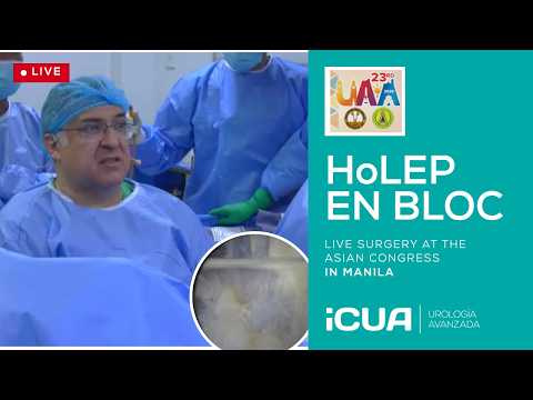
Live En Bloc HoLEP during the Asian Congress in Manila
Dr. Fernando Gómez Sancha · ICUA
Dr. Fernando Gómez Sancha · YouTube
Language: English. YouTube audio metadata.
Surgical content for professionals. Contains footage of an operation.
Playback connects to YouTube. Your search query is not sent to the player.
About this video
A live en-bloc HoLEP recorded during the Urological Association of Asia congress in Manila. The session is intended for urologists and trainees interested in surgical technique and operative judgement. The description identifies the Magneto laser, Richard Wolf instruments and Piranha morcellator, and acknowledges the companies that supported the session.
Documented details and sources
Only details explicitly stated in the sources. Missing information does not mean it did not occur.
- En bloc
Live En Bloc HoLEP during the Asian Congress in Manila
- MAGNETO
I would also like to thank the sponsors who supported my participation: Quanta System, for the Magneto laser, and Richard Wolf, for the surgical instruments and Piranha morcellator.
Source checked: 2026-10-06.
Available transcript · English
Texto y tiempos de la pista publicada por el canal. Puede contener errores; no se utiliza para inferir atributos clínicos.
They may contain transcription or translation errors; check the explanation in the video.
- you use the jelly it would be nice to have jelly on a inside the syringe we inject it Bravo Bravo now let's get the instrument first okay so this is the outer sheath this is the working elements so take it out and let's put the new one very nice because it filters the water so the camera doesn't get wet that's old that's okay now where is the external sheath you have it there and
- then the optical optical optic the 30-degree lens where is the lens here is it dry Bravo 30 30 is 30 no is 12 okay 30 clean it dried right up it needs to be completely dry completely dry
- otherwise the camera will get wet okay 30 30 okay so now for the camera I need the sheath the the , yeah no this which cable are we using this one okay okay okay so let me have this one so can you throw the camera in here now you take this this bit okay and we pass it under the leg huh like that okay and now you have to help me you see there is a where is it I don't know I don't know I don't know I don't know I don't know I don't know I don't know I don't know I don't know me it it's where is the hole here yeah there is a little hole so you need to pull the ears a little bit to allow me to put
- this inside so elevate and pull no just through here to pull you see if we can so you're like that okay that it's pull here so here like that okay that's it that says this is already protecting it that says this is already protecting the camera hopefully it will work now we have a cable please let's let's horse of the black source I CFR to wrap it in fitting a four in here please so so that is no this is uh just to connect here it's okay do you think this connects here this is very strange let's see now it's fixed okay very good white balance please oh okay who is doing the white balance and focus a little bit would it be possible to put the screen a little bit a little bit more
- into the yeah where's the white balance huh it's elusive general presets white balance okay okay good so we're waiting for the connection no this one's for the outflow i don't use outflow i do you have a clip uh so i can clip it here to my uh any any forceps yeah so do we know how are we in terms of the connection can you ask if they're ready or not so they are listening they're listening to us they're waiting for us to start
- and are the moderators ready because it would be nice to be able to speak to them um do we have a clip uh just grab it here yeah okay be careful with my balls okay all right okay the fiber i will ask for it in a moment let's put the jelly gel here inside the urethra so they tell me that people are already watching is it correct just inject it into the urethra yeah yeah yeah okay okay this is correct can we start or five minutes five minutes five minutes okay that's what i wanted to know you see at the beginning there's a little bit of resistance
- so we're going to use the otis for a moment huh do you have it ready do you know how it works just show me show me the otis and i will okay so this is the otis it has a blade you see the blade yeah this one and uh it has this possibility to open so you open it in the urethra and then you pull the blade and it will do a little cut at 12 o'clock very but you see that after that we go in and the only problem is you you can damage the urethra when you put it in so you need to be very careful you know when you put it in don't don't do any force just just a little bit like that okay so we do like that open until it's
- contacting you see it's not even fixed too much and then we do a little cut this is usually enough there's a little bit of blood coming out of the urethra which is quite normal and then you see the little cut it only cuts the the tight area you see let's see how we get in there it's going going in quite well so yeah camera let's try to focus i'm looking at that screen even better than this one this one is too too close it's possible to push back the focus there we are going in gently as i said before
- the surgeon must have the light the hands of a lady so it's a big prostate you you can tell because there's the veru is far away you see and the sphincter is here so there are like two two centimeters maybe of apical apical growth the the the adenoma is pushing the the sphincter far away from the veru so the veru is not a reliable a reliable uh landmark no so let's open that flow let's go in carefully it's a relatively large prostate okay not so small for for the residents you see you can you can see a surgeon manipulate an endoscope and know
- very well if he's an expert or not basically because the movement has to be very slowly very slow you see many times when i see residents it's more like this this is terrible okay so if you are manipulating the scope remember to use it very very carefully so you move you know from one side to the other and keep keep the keep the slow motion movement no okay so i will be ready to start we have the fiber thank you but we need to go from the from the audience are we recording this yes no so that's the fiber that's a fiber for for the operation we will we will try to focus on the fiber okay because this is where this is
- the distance we're going to be manipulating we're going to be using say again it's okay to start but i not i'm not listening to the moderators okay so when we come out you see as you come out as you come out you will see the sphincter closing at some stage you see that we see so this is the limit so we see nothing should it just start or or are they going to be speaking to us can you wait so we can't see you see this is the limit of the sphincter you see this is the lateral lobe lateral lobe and it comes all the way down to here here's where the sphincter starts you see
- this is sphincter has a horseshoe let's say anatomy so we want to protect of course especially the upper part it doesn't matter if you go down a little bit here that's very unlikely to cause much much trouble one of the problems of working at this level is that the sphincter closes over the draining holes from the from the you see there's stagnation of the water if i go inside and and if you look at here sometimes when you go inside then you see that it's outflow you see so there's no need to say yes good morning doctor sanja hey i can hear you good morning yeah we are already live you're already live okay so this this is uh i i guess the case has been presented already so we're going to kick off with the operation you can see that this man has a very long frenulum this is a varro and it goes down a lot and when we come out
- you see we can see the sphincter closing here you see sphincter now so in the traditional teaching of TURP you shouldn't resect below the veru but all of this is adenoma okay so to start and to have a good good landmark I'm going to mark in both sides of of the of the sphincter you see both sides of on the and I want to go very very close to to the edge between the sphincter and the adenoma okay this is what I call the white line and I think the white line is we have this interference is this normal this interference you see that the image when I fire the laser is getting strange no it's looking bad Dr. Sanchez this is a Kazumi Taguchi from UAB Urology yes hello you explain what is your laser setting right now ah settings are used [250*] [250*] for for dissection and that's going to be with
- the virtual basket pulse modulation setting because it's a laser laser laser laser laser laser laser laser laser laser laser laser laser laser laser it's a it's excellent but I use the classic setting [250*] huh and then for coagulation we're going to see magneto magneto as you know is a is a setting that mimics the long pulse of the TFL laser and it's going to give us excellent hemostasis you see magneto only allows you to use [32*] watts of power so it's not it's a very good setting for stones and for soft tissue like bladder tumors you know when you want to do bladder tumors but uh of course uh not so good for enucleation some people are struggling to do enucleation with magneto but I think it's not worth it it would be a very low power setting okay so this is the white line is going to be our reference of where is the sphincter for the operation see the sphincter is here it's perfectly
- safe and now we're going to go to the floor of the of the paracollicular you see here we're going to go down and we're going to start uh exploring a little bit this area letting the bubble find the way to try to find the entry to the to the plane you see to the anatomical plane between um adenoma and capsule all right so this little movement is going to start opening up the the plane what we want to do is we want to get to the paracollicular space here and get an initiation of the of the plane in both sides so this is one side you see here we entered now I'm going to go to the other side here and I will do the same I will enter carefully and you see all my movements are like in slow motion huh so you see the operation uh has to be very controlled let me focus a little bit better but it's a little bit
- uh bothersome to have this interference with the camera no sometimes the very good cameras are very probably they can see some of the infrared lights flashes that we cannot see and it's difficult for the cameras to interpret these changes in intensity no and so this is a common problem for 4K cameras for example but there we go you see I'm doing the same maneuver on the other side I'm not obsessed with recognizing the anatomical plane and I just want to enter this paracollicular space and then I will try to connect both spaces cutting over the veru and uh the way I think on the on the prostate I I think the prostate is a boxy structure it's like a box okay so I'm going to discuss during the operation four
- planes we have the posterior plane we have two lateral planes and we have an anterior plane okay it sounds very silly because we know that the prostate is more pseudo-spheric but I think it helps a lot to uh systematize the operation like that because when you are feeling lost at least you can say where am I posterior lateral or anterior no so that's that's a beginning so here as you can see I am keeping this line of dissection also I will talk about the line of dissection and about the plane okay this is the plane this is the line of dissection and for me I like to keep very uh very um organized lines of dissection because when you have a good line of dissection you can be very well oriented and that's why I do ample movements you see I don't
- do short dissection like this because if you do that you see we're getting deeper here but here we're not so deep and here we're not so deep so what I want is to do long movements because the long movements are going to give me uh a line of dissection that is very structured very continuous it's more or less at the same level let's say and it will let me organize the the operation very well so here we are moving from side to side sometimes we can move we can dissect the posterior plane a great deal for some reason some cases allow you to to progress very far even close to the to the bladder neck and some other places too much access if you think about it we are under the the adenoma this would be the urethra we are under the adenoma and there is a fixation here you see the the prostate still fixated
- there so basically we do what we can I don't want to force anything and I have to tell you about my philosophy of enucleation okay so for me an enucleation is a yes yes yes go ahead just just some questions Do you use active suction here, in this case, or just passive suction? No, I use gravity. I use gravity outflow. Yeah, I don't connect a tube to the outflow. And what kind of laser fiber is this? It's a 550. 550, yes, yes. It's pretty standard setup. Is it unstable, the laser fiber? Yes, it's a little bit shaky. It's a little bit shaky, but because I'm using the insert I used to use for the green light fiber, you know, when I was doing... I see, I see. So, it's a little bit shaky, but nonetheless, the amount of shake is very limited, so I can still be pretty precise, okay?
- So, here we are. Yeah. Let me tell you about the philosophy of enucleation, what I am reflecting, you know. I spend a lot of time every week doing cases and... Let me, let me philosophize, make some philosophical thoughts about enucleation. Okay, so, basically, what we do in enucleation is we release, we release the capsule from the adenoma, okay? But the idea is that when we release the capsule from the adenoma, we gain a little bit of mobility, okay? The adenoma starts moving a little bit independent from the capsule. And this mobility is going to give us... Access to places where we couldn't access before, okay? So, basically, the operation is going to be a succession of movements that are, let's say, oriented to get more liberation of the plane, but it's going to be very strategic in the sense that we want to do things in a way that stretch the sphincter as least as possible.
- Here, I was doing a little bit of an access incision, and now I'm going to start the lateral dissection. You see, now the movement is more... More up and down, let's see. Up and down, and I'm going to try to connect the line of dissection that I'm developing now with the previous line of dissection, okay? Because we want to develop a circumferential line of dissection around the adenoma. So, you're going here for an en bloc technique? Yes, the idea is... You're always using en bloc techniques? Yes. There's never... I mean, you cannot say... You're always in medicine, no? Yeah, of course. This is my... This is my... Just for thinking for beginners... Yes. Is it useful to learn a two- or three-lobe technique, or directly start with an en bloc technique?
- Well, you know, en bloc is getting more popular nowadays. So, I would say that you have to learn what your teacher... What your mentor can teach you, you know? Yeah, I teach en bloc, and I find that residents and people who learn with me learn this process very, very easily. But, of course, if your teacher, your mentor knows how to do three-lobe, it doesn't make sense to say, teach me en bloc. So, I think... You have to learn from a mentor, and a mentor has to know the anatomy once you know the business. How to dissect. Then, of course, you're going to be able to experiment with other techniques and other options. Okay? So, you see, this is the posterior line that I was carrying before, and now I'm coming up.
- But, you see, here, I'm very sensitive to difficulty to go up, basically because I don't want to force the sphincter anteriorly. You see, there are attachments here from the adenoma to the sphincter. You see that? And I want to release the apex before we can move up with our dissection. So, that's why I do these incisions here. I'm not looking for the plane. Actually, many times we cut on the adenoma. But, you see, when we cut on the adenoma, basically, we're establishing a separation between the sphincter and the adenoma here. You see? So, that gives us a little bit of space and separation. And then we will go to look for the proper plane, which is different. So, here's an incision. You see, it goes more into the interface between capsular and adenoma. And now we're going to bring this lateral line a little bit further up. The general objective is going to be to go from 6 o'clock here at the lateral lobe all the way up towards the 12 o'clock.
- You see that I move pretty slowly. And also, I am always concentrated on trying to use the energy to have a soft effect on the tissue. All right? If you control the effect of the laser, you can have a soft dissection effect. And I will never have bad perforations because, you see, the way I use the energy is very soft. It's very difficult to cut through the capsule when the distance you choose is a very careful distance. Okay? So, basically, when we are moving in this plane, for example, towards the bladder neck, you see, when I'm dissecting the lateral lobe, this is it. You want to drive a car. You want to stay in the road. If the car gets too close to the outer part of the road, you will have to change your direction a little bit to stay in the road. So, here, what I'm doing is exploring as we go.
- You see the quality of the plane. And I look on the left side. I have fibers. It looks very capsular. On the right side, it looks like adenoma. So, I'm okay. But in the event that I am in my direction. It's going a little bit deep into the capsule. I will be in time to change the direction before I perforate. Okay? And also. Yes, go ahead. Go ahead. Do you apply any amount of force or you just let the laser dissect the plane for you? I don't do any force. What I do is a little bit of contact. You see, if I work like this, for example, trying to dissect like that, I have no control of the effect. Get closer to the line of dissecting. You see, I'm not pushing. I'm not doing force, but I'm letting the tip of the endoscope is generating some traction and countertraction, you see, between adenoma and capsule. And that is going to help me a lot to control the effects.
- Okay. So, we went up more. Okay. And here again, I'm coming back to the area of the sphincter because I want to continue doing another cut here below the sphincter because we want to go below the sphincter over the adenoma. Okay. So, I'm going to go below the adenoma. Initially, this cut will give me some access. I call them access cuts. And then I'm going to try to continue. Let's see if we control these little vessels. Try to continue going up. You see, to try to detach the adenoma. I don't want to leave any adenoma in situ. Okay. So, but the principle is that we don't pull the sphincter. If you remember the encircling maneuver, the encircling maneuver that was done before in the classic technique, you would go from 6 o'clock to 12 o'clock, but you would never go back to look at the sphincter. And what happened is that you would see at the end, you would see this 12 o'clock flap. And I call it the infamous flap, you know, because basically what was happening is
- that your endoscope was tearing the mucosa of the sphincter. And this formed this 12 o'clock flap. And that's accounted for a lot of these postoperative that was observed with the classic hole. And the objective here is to do a very careful early apical release. Okay. It's not an immediate apical release, but you see, what I'm doing is trying to accompany. You see, I'm following this white line that I marked to know that I'm in the right depth. Here we are reaching the 12 o'clock region. You see. So basically, the idea is that we do a very careful separation of the sphincter and the adenoma. And normally, we're going to see at the end of the operation. Let me finish and we're going to see at the end of the operation that the sphincter keeps the mucosa. Okay.
- And that is going to reduce a lot the incidence of stress incontinence. Go ahead. Go ahead. So just a question. What means early? Within one minute, five minutes, ten minutes. It's usually it's going to be usually five or six. So into the operation. But you see, following the philosophy that I told you, the idea is that we want to generate a comfortable mobility of the adenoma. You see a constant mobility enough so that we can reach the 12 o'clock without doing a lot of traction. And here we are starting the anterior plane. You see now the direction of the plane is changing. And it's it's. Also giving me a way to to talk about another very important concept. You see, when we are
- doing an end block technique, we have the opportunity of support. Let's say the weight of the endoscope over the adenoma. Okay. So when we come to this is becoming a little bit bothersome here. Sorry, let me change it so it doesn't interfere with me. Okay. There we are. So. The idea is that once I'm on top of the adenoma, my endoscope can do a little bit of weight. I put some weight on the adenoma and that's going to mobilize the prostate inside the pelvis. You know, the prostate is fixed to the pubis by the puboprostatic ligaments and of course, by the urethra. But the rest of the prostate inside the pelvis, it's quite mobile. So the moment I come on top, you see my endoscope is bringing the prostate prostate a little bit down in the pelvis, which means that we don't have to angle the endoscope so much despite being in the anterior part.
- Okay. When you when you try to do a 12 o'clock incision, normally you have to tilt the adenoma enormously. Okay. So here you see this is anterior. This is anterior. Now this is the 12 o'clock area. Okay. So we could do a little, you know, again, access cuts. And we will leave this here. And now we will do exactly the same from the other side. So we can meet at 12 later on. Okay. So now this is the white line. You see how comfortably I can see where is the limit between sphincter and adenoma. I will deepen. This is you see the mucosa of the sphincter. I can deepen the incision a little bit again to try to find the entry here into the lateral plane. Okay. So we will start doing developing a little bit of the posterior aspect of the lateral plane. Let's see if we can slowly investigate. Now, one of the beautiful aspects of enucleation is that
- you need to discover the anatomy as you go. You know, it's similar to when you dissect the kidney pedicle. You initially don't know exactly where things are. And you have to discover here is the same. We are exploring the plane and we are finding a way. So this is the posterior line that we did. Now I'm coming up. You see coming up and down to try to start the posterior part of the lateral line. Let me see. This is bothering me a little bit. Okay, there we go. So up and down again. And then another detail is that, as you can see, I have developed the skill. And this is something you have to do when you do your whole lips. I always think about the people from the sports business. You know, football player or basketball player is not only playing basketball for training, they go running, you know,
- because they want to maximize their skills to be good basketball players. So we have to do the same. We have to not only operate one case and another case, but in every case we do, we have to try to improve our technical skill. So, for example, some people do like this. They do a little bit of dissection. Then they stop for some reason and then they go back and then they continue with the dissection. But you see, what I do instead is when I have to go back, I continue with a dissection. And this is making me save a lot of operative time. Okay. So you see, when you move slowly, when you use the energy very carefully so that you can perfectly see what you're doing, the operation looks how do you say looks slow, but is fast. You know, because we are moving forward and we are advancing.
- So every minute of the operation, we get some we get some some advancement. Okay. So keep the pausing time to the minimum. And here is the same principle. I want to dissect the lateral plane. I want to dissect the some reason I'm not very comfortable with the drape. Okay. It's okay. It's okay. You see, we want to progress towards 12 o'clock, but I will do it very slowly and very carefully. I don't want to do and I don't want to notice any force. If you look at my hands, most of the time I'm holding the camera with my fingertips. So I'm not doing any force. I don't need to push. I don't need to. I've seen people bend, you know, the instruments against the patient. And I think we should try to avoid that kind of leverage. So, again, I feel some resistance up here. You see, so I come down again and I have to go towards 12 o'clock.
- Following the white line. So I know that the sphincter is perfectly safe here. This is the 12 o'clock and I want to go over the adenoma under the sphincter. Okay. We are we're trying to get there up there millimeter by millimeter. We want to do one millimeter at a time. Let's see if I can finally conquer and finalize the early apical release. I said it's not an immediate apical release because you see many times the edge of the adenoma anteriorly could be one or two centimeters above the sphincter, you know. So when you want to conquer this anterior part, you need to have enough mobilization so that the adenoma lets you get there without having to do a lot of force. So you explained before we always
- normally starting from the posterior dissection and going to bilateral and then finally going into the anterior prior space. But is there any specific situation you do differently? You see, I have been very, very lucky that I was going to I was invited to operate in Bulgaria. They have this small clinic and but they are operating there for 17 years. So I go four days of the month and we do 25 or 30 cases every month in four days. So this is a very, very hard work. And it's also an opportunity to try new things, you know, and also as a teaching center, you know, because many people want to go there because they can see many operations in a short time and get a very good teaching experience.
- Of course, these people ask you questions, you know, like you're doing now. And and they make you think and you have to be able to explain things. And so I have tried many, many, many options. I've tried many. Strategies and at the end, I think that this this is a universal or nearly universal strategy. So here you see, we have the plane in one side. These are the 12 o'clock fibers. You see, not the normal has a little bit because we released some of the anterior parts, you see, now the other normal is fully mobile. So basically what I have to do is to come here at the apex and do a cut that goes from point A here. You know. Side, we reach to point B to be the other side, the plane can focus and there we are. So this is the early apical release.
- This is where it ends. OK. But it's also a careful release, not just going there. Some are recognizing first technique. So they try to enter the plane at 12 o'clock as the starting point. But I think it's going to cause a lot of traction against the sphincter. I recognize that this initial phase, there's traction on the sphincter, but I think the slow movements, the slow movements allow the sphincter to adapt quite well. You know, if you are doing very fast movements or very repetitive movements from side to side or things like that, you're going to stretch the sphincter maybe without giving it time to adapt. And we know. And if we look at the other sphincter we have in our bodies, you know, the anal sphincter, we know that it gives a lot. It gives a lot. It dilates a lot. If you do it carefully, you know.
- So general surgeons just so just one comment. Yes, because you have to think about the times when we do open surgery. Yes. So if you do a Freyer's procedure. Yes. Procedure. We are doing it with maximum traction and we see nothing. And finally, patients were not incontinent. So I'm not sure whether this concept of early apical release really is the reason for early incontinence or not. I think it's the experience of the surgeon. So and I think we have some data of learning curves. It's more or less the problem. When when you're not so experienced that you're doing a lot of unnecessary movements at the level of the force at the level of the sphincter and not
- and not the early apical release, I think. Well, we don't know, and there are things that we will never know. So, right. Sadly, sadly, with the enucleation, the quality of the studies. We read and we do is it's very, very, very low in the sense that and when you have to rely on expert opinion, you know, this is the lower, lower level of evidence available now, so I always encourage people to be. How do you say don't take what I say for as the truth. You know, just make a mental note and listen to other experts and then see what you experience yourself. You know, because because for me, the early apical release means that you're going to be careful. So, you know, if we go down now to the sphincter, if we go out, you see,
- probably, probably we will see a sphincter that has still mucosa recovering. You see, there's mucosa on the lateral wall. There's mucosa and clearly we don't see the 12 o'clock flap. OK. OK. And this is probably one of the important factors. Then there was a paper from Mario Sofer from Israel. He was saying that because he was doing a ureteroscopy on a patient and HoLEP at the same time. So he had the he had the X-ray and he looked at the position of the sphincter. So positioning the endoscope near the sphincter and after the sphincter. And he marked with some staples. He marked the drape. To see the location of the sphincter. And then he would take a shot with the X-ray during the lateral dissection, for example, and he could see that there was a lot of lateral displacement of the prostate
- during the enucleation. But here is where I think the en bloc makes an advantage. You see here I'm working on the lateral lateral side, but my endoscope is pushing a little bit the adenoma towards the left, you see. So maybe when I'm working here, the prostate, I am capitalizing the mobility of the prostate inside the pelvis to try to reduce the lateral traction. You know what I mean? It's it's very interesting. And when you do it lobe by lobe, maybe you lose this this possibility to to compress a little bit the adenoma and to push the adenoma, the prostate inside the pelvis, mobilize the prostate inside the pelvis. This is the entry into the bladder. You see, I'm on top of the anterior here, and if you look at my scope, I don't have a massive angulation. So I can you see I can bring the prostate down in the pelvis and I don't need to to to to to to do this very aggressive movements.
- So as you said, there's no probably no single reason why we get so low incontinence rates, less than one percent after three months, you know, it is it is virtually. I don't think you can you can improve that, you know, I. Because we treat patients with a lot of heterogeneity of situations, old patients, you know, Parkinson's patients, big prostates, long term catheterization patients, I don't know. So I think it's having zero incontinence is probably not possible, but we are very close to zero after after three months. And and that's probably because of a number of things. One of them might be the contribution of the early apical release and the careful dissection of the. The sphincter and maybe the other reasons are the preservation of the mucosa, the let's say avoidance of excessive traction when we do
- this mobilization of the prostate inside the pelvis. So let's see where is the [ureteric orifice?]. Now, you know, I don't do stone, so for me it's very difficult to find you. So it should be somewhere probably. Far away from us, so we are quite happy that we not we're not going to damage the [ureteric orifice?] today, hopefully. And now you see, basically, when I when I was at the at the sphincter, I was doing six o'clock to twelve o'clock and now we are going twelve o'clock to six o'clock, OK, so basically the the structure of this operation is very simple. We do posterior dissection first, then we do apex going from six to twelve in one side, from six to twelve on the other side. Then we go [on ...?] mostly to get to the bladder. And then we do this twelve to six dissection.
- Of course, I have to go back and forth. But the philosophy is that I'm going to come from twelve towards six, you know, in both sides to try to do the posterior dissection here. You see that the camera is always aligned with the line of dissection. So we keep half of the image is probably adenoma and the other half of the image is probably capsule. So we can see very well. I can see now. You're using the straight camera, not the pendular camera. Yes, I prefer I prefer the straight camera. Yeah. And I prefer not to use the trigger. You see, some most companies have adapted no resectoscopes to to allow for enucleation. I know that Peter Gilling started using the fiber like I'm doing. He used a catheter to stabilize the fiber, but he was handling it with his own hand.
- And then I think it was Kunz from Germany who decided to invent this trigger. But I always say that this is like, you know, when they invented TV. The first thing that they did was to film the radio, you know, because we humans are not very able to we're quite able to improve what we do slowly. So with small changes, we improve what we do. But we're not so good at inventing totally new things. No. So I think to me, the the the trigger first, you see that my movements, even when my fiber is shaking, are very precise because it's a simple movement. If you use a loop, mostly you have to do this kind of this kind of movement. When you go up, when you go down, you see it's rotational. And then there is the up and down movement on this at the same time.
- But I'm doing this very simple, very simple movement. So it is much more simple. And I think probably despite a surgeon can compensate for I mean, if a good surgeon can compensate for a bad instrument many times. But but I like to work with my fingers on the on the fiber. It gives me a lot of sensitivity. And you see, I can work with very simple, very slow movements. And if you keep the energy because, you know, the energy many people ask me about the settings and I tell them, you see, if I'm too far with the same settings, there's no effect. So I need to get closer and maybe I'm coagulating, I'm not dissecting, you see, but then when I get even closer, then I get some dissection effect. So I always tell the story about the you know, someone walking in Paris and he sees this artist in the street trying to draw a portrait, you know, and they ask that they ask the artist and they say, oh, you have a nice pencil,
- you know, because the the the portrait is so beautiful. And then he gives you the the pencil and say, try yourself. And then the portrait is not beautiful at all, you know, because, of course, the painter, the artist has has understood the different kinds of effects he can get on the paper when he's drawing, you know, and the same way when you use a laser fiber, you have to be able to understand the different kinds of effects you can get with the fiber. So in some moments we will do contact because we want to be very aggressive cutting. And this is, for example, when I am close to the bladder neck, if I want to cut the bladder neck, I might be doing contact, you see, to get a more disruptive cutting and to more efficient cutting. But when I am here down here, for example, you know, this angle now the fiber is mostly perpendicular to the to the to the capsule. So I need to to to tailor how I am going to fire.
- I'm not going to fire down here because if I fire down here, I'm going deeper in the capsule. I'm going to fire up here, you see. So as a surgeon, you have to use the fiber in a way to get the effect you want from the energy. So the settings are only determining how the energy comes out from the fiber. And the surgeon is responsible. I'm trying to see if I can get an access here. You see, this is now bladder neck almost below the adenoma. So I want to free this because you see, the next step is going to be interesting, too. Some people tell us en bloc, you know, the prostate is sometimes wider than the bladder neck. That's why en bloc doesn't is not good for people who don't know much about this, about en bloc or have no experience. But once you have this kind of liberation, you can lift the prostate, put it there,
- leave it there on the bladder neck, and then when you push a little bit here, the the adenoma is going to come out. It's going to rotate. You see. So this is now the prostatic urethra and this is the bladder neck. So you see that lobe has gone into the bladder. And now we have much better view here of what is remaining. You see, so we can then finalize and we have access to the most posterior and lateral aspects. Here, one of the dangers of pushing too much is that you can pull from the other side and the capsule can be very thin here. So that's why. I'm getting my fiber very close to the adenoma to try to lift the capsule in place. And there we go. We are nearly finishing now. You see, we followed a structured approach. Always the same. When you follow this approach all the time becomes very familiar. And even when the plane is not so nice
- to recognize or when we do cancer patients or you get used to the steps, you get used to the anatomy and to me is a very very strategic way of doing this operation. Another good thing of the block technique is that for the majority of the operation, you are irrigating a very small space. You see, so if you do an incisional technique, if you do an incisional technique, you have to cut the bladder neck and there will be some bleeding that blood will enter the bladder and then you will have to exchange 500 milliliters to to to be able to see, you know. But when you are doing an end block technique, maybe the amount of water between the capsule and the adenoma is 10 or 20 milliliters, which means that probably to exchange that water takes seconds. That's why you get very good visibility throughout throughout the operation.
- Here we are. This is the end. This is the snooker maneuver. You see, when we push the adenoma into the bladder and now I'm going to be careful to. Do very good hemostasis before we move on to morcellation. No, it went well. It went well. Yeah, this is amazing. So there is some you really are gravity of the order. I got difference between a novice surgeon and someone like you for experts. Well, you know, one thing that one thing that has stricken me very hard is that incredibly, incredibly with. With all the video. Available online, you know, YouTube videos and stuff. I'm seeing people doing HoLEP and I am very impressed by the quality of their work. You know what I mean? So it took me more than 10000 cases to to to to to get this experience.
- But when I see my residents and and people who work with me and and people who I teach, you know how fast they become really, really good at at doing HoLEP. Huh? I have to say, I was both happy, of course, for my pupils and for. But also, I felt like, wow, this this if you allow me, these fuckers, they they have they have the advantage, no off of being taught properly. And you understand what I'm saying? No, I'm mostly happy. I have to say, but the pupil is always better than the master. Yeah, this is the idea. This is the idea. They have to get much better than the master. And here, let's see. You see, now when I try to reach the anterior part, it's getting a little bit more difficult for me because because what happens with the water as we have water? Do you prefer a total decompression at that point before most elation?
- No, no. Normally, every leader normally no, because despite the quality of the hemostasis that we do, sometimes there will be some venous openings, small veins or and then when you decompress completely, the bleeding will start. And then I think it complicates the a little bit. I'm not used to to do that. Do you do that yourself? Not regularly. No, but after most elation, after most elation. Yeah, yeah, yeah. I think it's we do a very fast change of instruments trying to keep the pressure. And, you know, one of the one of the problems with more selection is exactly that we have this fossa, which is sometimes full of little venous openings that are not bleeding because we have some pressure now
- into the bladder and. So some people, let's say, stop the inflow. So if I stop the inflow now, for example, I might be able to see better. I don't know where the where the bleeding is coming from now. But so the problem with more selection is that we're going to be sucking water. Also, there will be some water going in. But if we suck a little bit of water out of the bladder, you know how the bladder works, if you do your dynamics, you know that the bladder fills without any rise in pressure until it's very full. So basically. At the end of filling the last 50 CCS that you input in the bladder are going to cause the increase in pressure, which means that if you empty the bladder 50 CCS, the pressure is going to go down to zero near zero again.
- No. So during more selection, if you manage to keep the bladder full, then all these little veins will not bother you. But if you suck too much water, then the bleeding starts. So it's a little bit. It's challenging sometimes. You see, because even with modern lasers and magnetos and virtual baskets and everything, sometimes during the dissection, we go a little bit deeper in some areas. We find these little veins. So you see, it's a it's a challenge. So what is your experience of en bloc techniques, let's say 300 CC in en bloc? Yes. And then the modulation process, because in my experience, sometimes. So let's say I've done.
- I have done or I do five to seven cases, let's say 300 grams per year. But in these cases, if you do it on block, you have problems with modulation because sometimes you need one hour for, let's say, 30 grams or so. If you. The large ones. I do, I'm looking in the large ones, too. And it's interesting because because, you know, time the modulation process. Yeah. Yeah. You know that Karin Lerich looked it up, you know, if en bloc let's say change the modulation time or not. And they found that there were not significant differences. They haven't published it. Yeah, I think there's a lot of variability. The large ones, let's say 300 gram en bloc and then you use the modulator.
- So my experience in the really large ones, you have problems with modulation because the bladder volume is too small for because you need some space for some mobility. Yeah, yeah. Some mobility. Yeah, it's it's a possibility. I don't feel the same way. But of course, there are cases and cases. Sometimes the bigger prostates are the softer ones, you know, and they mostly reasonably well, sometimes not. No. So this is the problem. What we do and what we think is not not so scientific. It's much more based on our experience and perceptions. No. And. Yeah, but of course, splitting the prostate, for example, once you have done an en bloc, it's it's very simple. It's very fast. So you could as well do that if you feel that
- you can split the prostate before, you know, probably mobilizing it or. OK, so I think we are getting there more or less. Let's see. Usually there are bleeders apically. Let's we don't want this to be going while we morselage. And yeah, sometimes you see I was going relatively slowly. Yeah, but sometimes. The really difficult aspects of enucleation is to find the right tempo, meaning that, you know, the composers of the past. When they wrote their music, they had to give an indication. To the directors of how fast they need to to play the music. No. So they had this Italian terms, you know, Allegro, Allegro, Manon, Troppo, et cetera. And here we leave a little nodule. So I think. I think this tempo is one of the most difficult things to to master with
- enucleation, meaning that if you go too fast, sometimes the time you gain by going fast is lost later on because you have to do more hemostasis. And sometimes if you go too slow, of course, you go too slow. So. So, yeah, finding I mean, the ideal situation would be where you finish your enucleation and then you have to do minimal check on on hemostasis. No, that would be, to me, the most time effective, let's say, balance between, well, the tempo, the tempo you choose to use. No, let's see. There's a vessel down here which might cause trouble. You need to check the Apex here and that one, of course. Yeah, yeah, yeah, yeah. These things you learn painfully, you know. But yeah, I think I think I really think there is a massive.
- Change, you know, in the way enucleation is being perceived now, most centers want to do enucleation, I think most centers are doing in many countries, I think France is very advanced in. Doing all of our enucleation, I don't know, Germany, Spain is quite it's quite advanced in Germany. We had. Three thousand procedures out of 80,000 procedures. OK. Until 2018. And now we have 20,000 enucleations. All right. So it's one fourth. It's 25 percent. No. And in Brazil, in Brazil, they went from nothing to 25 percent of cases are done by HoLEP in Brazil in five years. So there is a tremendous interest and tremendous uptake. And because I think that teaching has been has been simplified.
- The videos are helping. And now there is more people who can do HoLEP. So they can mentor, of course, more for more centers. Let's change technique is better fiber because you stay in the same plane. So if you do it in two or three low lobes, then you open different planes and you have to to to to find connect them. Clean this. We need to help me with the microscope. That's not so easy for the beginning. Clean. Clean. This with with the white goes white goes clean. This is also the difference of the side also. It's a laser technology. Yeah, it does, you know, provide us a lot of advantage. And you know, it's like in the day we have to use a very low power laser. That's how we did mechanical dissection with like a laser dissection. But now we don't have to do so.
- It's venturing. So I'm going to close that. For. Give it to you. Take it out. And now we change. OK, there we are. More later, please. Second inflow. Yeah, normally at home I don't use a second inflow, but for life cases I do. Because I think maybe the instrument is a little bit different. So here we are. Now we have opened the inflow. Just leave it like that. Leave it like that. So now let's focus. We have to focus a little bit differently. The bladder is is certainly filling up. I think we get the right focus. We see the adenoma and we kick off with morcellation. OK, so as I said, the problem with morcellation is that there is a decay in the visibility during morcellation. When we start, we have good visibility. What? Just a question. What's your setting? 750. So it's a one thousand five hundred.
- So it will rotate. 750 to each side. And I use two hertz, you know, hertz. This is a setting in the Piranha morcellator. But basically a hertz is one cycle per minute and a cycle means sorry per second. And a cycle means moving right and left. OK, so here it's two hertz, which means that it will go right, left, right, left in one second. So zero point you perform upside down. Yes, yes, I love it. I love it, too. It's not so good at the beginning when the adenoma is very large. But I, I, I will tell you what I what I think about it in a moment when we do it. More salating the fossa or the bladder. It depends. I start in the bladder because bringing back a big adenoma into the fossa is not so easy. So once the size has been diminished, if you still have very good visibility
- in the fossa, then you can come to the fossa and morcellate. Comfortably. So the idea is that. There is no time to lose with more solution, OK, because the visibility is going to decay. Most of the cases you will get worsening visibility over time. So we have a window of opportunity of about 15, 20 minutes where the visibility still holds. Sometimes we're lucky, sometimes we're unlucky and it goes faster. So the idea is that you don't want to lose time. So the same way that I was telling you before, even when you move slowly, keep the pausing time to a minimum. In morcellation, you have to keep the pausing time to a minimum. OK, so you don't want to go and check the value for nothing. There's no use in that. And you should move, let's say, the least possible. So for me, how do you know that morcellation is safe? OK, one thing is that I will never
- keep the morcellator sucking water on the air like this. If you do that in about. I don't know. Very short time, the bladder will be completely empty and then there is risk. And of course, we said before, if you empty the bladder, the pressure goes down, the bleeding starts. OK, so what I what I have developed is this idea of the black triangles. You see many people morcellate and they keep the morcellator very close to the to the to the window, to the lens, because they are very afraid of causing a bladder lesion. You see. But when you do that, the whole screen is covered with tissue. So you have no orientation of where you are. So I advise to put the blade a little bit more inside. You see now that the adenoma mainly stays anterior in the image and in the posterior part of the image, in the lower part of the image, we see the blade and in both sides of the blade we see a black triangle.
- Why is it black? Well, because the light of the endoscope is reflecting on the adenoma. OK, so it doesn't reach the bladder very well. And it's also causing this adaptation to the luminosity. So many of these camera systems, when there is a lot of light, they reduce the sensitivity. So you get this black, let's say, black visibility, black triangles. Now we are in a not uniform. But you see, when we get closer to the bladder, the triangles become pink. So basically what I do is I capture the adenoma, lower my hands, get away from the bladder and hit the pedal. And then. If I see that the contact of the tissue on the morselator is good, that means that there's not so much water coming out because you see, it's the tissue filling the filling the mouth of the morselator avoids to suck too much water. OK, so I. The crucial step is to have a look at the irrigation fluid and to have a nurse
- who has a look at the irrigation fluid not running empty. Yeah. Of course, of course, that's that's super. Within within seconds, the bladder is empty. Yes. You know, that whole or an enucleation is is a teamwork and we can work. Well, how do I say that when you do a live surgery and you have lived that before, it's very stressful because you have to control everything in the operating room, the water coming in, if the change is going to be ready, if the instruments are ready. So you feel stressed. No, because you have to have all this concentration on everything that's going on. But when you have a good team and here I have to say the team is amazingly good. Let's let's try the upside down, because you see, there's another factor with well, let me finish with the team. I have so many things to say. The teamwork is fundamental because if you can rely on your team,
- then you as a surgeon can concentrate on your job and know that everybody is doing their job. OK, so. So that's why I encourage silence in the operating room. I encourage discipline. I think it's very important to have all the team present in the operation, which is not the same as having the team present in the operating room. Many times you have nurses. I think you're performing here very, very impressive surgery. Perfect. And in a way, games always different to being at home in your home. Are. And I think the pressure is always enormous to the surgeon. And I think here everything works really perfect in this. Now, the team, the team is very experienced. And of course, Karl Tan, I'm sure he's a tough, tough boss. Hey, what do you say? Can you tell us something about Karl to share in public?
- Probably not in the camera, not in the camera. OK, OK. Later, later, maybe. So this is the upside down morselation. This is this was, I think, Japanese idea. And it works really well. I think the mechanism is that the adenoma really wants to float up, you see. So when you're sucking it from below, you have to go against this tendency of the adenoma to float. And when you're sucking it from above, maybe it's like, you know, riding a bicycle down the road now rather than up off the road. So the adenoma, as you know, is a multinodular growth, benign growth. The adenoma has this stromal component and the glandular component. If you think about it, the glands are empty, empty or they're full of liquid. But basically it's like a sponge tissue. It's very soft. And there are some of the nodules are going to be true fibromas.
- When you take these nodules to the pathologist, he tells you this is all stroma. OK, it's a stromal nodule. So they're much harder to eat. So I always talk about the peach example. You know, when you eat a peach, you can pierce the meat of the peach with your teeth very easily. But when your teeth reach the bone, you know, the bone is so hard that you cannot pierce it. So the morselator behaves the same way. It eats the soft flesh, but it's very difficult to eat the harder flesh. All right. So at the end of the morselation, when you have the worst visibility, because the visibility has already decayed, you have a very tough nodule to morselate and it seems that it doesn't morselate very well. So what do you do? We call these the beach balls because sometimes they look like a like a beach ball. Well, one thing you can do is you can try to bring the adenoma inside the fossa. Because in the fossa, the tissue cannot escape so easily.
- Now, in the bladder, if you lose contact, the tissue will go away, sometimes far, and you have to capture it again. But now I am morselating inside the fossa. We were lucky to do a very good hemostasis. So you see, even when the tissue is a little bit hard, I can re-establish engagement very fast. OK, we need to change the bucket. So let's change. I'm going to stop the inflow for a moment. Thank you very much. So we're having fun today. It's very nice when everything goes well. And I have to say the anesthetic team is very nice and everybody here is very supportive. We had an excellent breakfast this morning. What about second breakfast? No, like like the hobbits. You ready? OK, so let's start again.
- You see, there is a little bit of blood starting to pull. Maybe the view is not as good as when we started. And then one one word of advice for those who are going to try to enucleate. There is a black legend about enucleation. People hear this you know, there is a lot of incontinence. I there are cases where horrible complications took place and the patient even died, you know, there are this is the black legend of enucleation. And and I'm not saying that these things didn't happen. OK, so these things can perfectly happen. But of course, when they happened, they happened because they operate, the surgeon didn't stop in time. All right. What does this mean? OK, so if you're more so if you are enucleating and you do a bad perforation, you can see the water flowing out. And you if you do an ultrasound, you see that there's water extravasation.
- But, you know, you just did a very bad perforation. So instead of continuing the operation, if you put a catheter, the patient is going to be OK, you know, when he wakes up, you tell him we came to a situation where it was not safe to continue. But if we go back in a couple of days, everything will be fine. We can finish the procedure and it will be all safe. If why do bladder perforations happen? OK, sometimes it's an accident and you had very good visibility. And I have to say, I did one like 16 years ago and then I never did another one. And so the surgeon has to know, especially when you're learning that if you encounter a situation that might be dangerous for the patient, stopping is the best alternative. You know, because if you have a bad perforation and you put a catheter, the perforation will close, you know, so nothing will happen, nothing bad.
- But if you keep trying to do the operation, the water will infiltrate the retroperitoneum. Maybe the pressure is too high and then the cava collapses and then the patient becomes eurodynamic, sorry, unstable and, you know, all sort of things. So in order to keep it safe, you have to have this this idea. There's no dishonor to stop an operation. I encourage it in my department and I do it myself. If I find that I cannot continue reasonably, then I will stop with a catheter and come back another day. I think this is a take home message for enucleation. You avoid the complications by being sensible. You see, this tissue is much harder. It's not it's not morcellating so fast. What else with morcellation? I think the black triangle trick is very good because you can do now because it's
- more and more harder here, you can reduce your speed. Yes. Sometimes it's more effective. Yes. If you go to 500 or 450. Yes, I would agree with that. That's a very good trick because you see, the morcellator relies on suction. If there is no suction, the tissue doesn't want to be morcellated. It will not go to meet the morcellator. So you need this suction in order to morcellate. And the way it works is that the tissue will is elastic. It's mostly elastic. So when you suck it into the mouth, it goes inside the mouth. It's similar to using a straw. You know, when you're having a drink, you take a straw, you put it against your fingertip and you suck and then the fingertip gets inside the straw. No, it's the same phenomenon. They see when the piece is very hard like this one, it's not so elastic.
- So you are sucking more water. So that's the that's the hard thing of morcellation, that in a moment when you have the worst visibility, because the tougher pieces are always at the end, you have to morcellate these hard pieces. So when you slow down the morcellation speed, basically what you're doing is you're giving more time to suction to increase the contact of the tissue with the mouth. And then it becomes more effective. We are we are more or less managing with the speed we have. Let's see. Eating the last fragments and I think we are nearly finished. Also, when you do an en bloc technique, you might finish with the piece you you're seeing and think that you finished. But you see, oh, surprise, there's another piece. And that's because the morcellator can split the adenoma for you.
- OK, so you should always check. If there is any other piece inside. And you should always look for a bladder diverticulus. Yes, yes, yes, yes, yes, definitely, definitely. Yeah, also, there are two situations that deserve a comment. When you have a stone, I prefer to do the stone first because with a normal mucosa, when you have a preserved mucosa, mucosa lining of the prostate and the bladder and everything, the stones can be extracted and you can get a totally clean, clean bladder. OK, if you have to if you leave the stone for the end, most likely there will be some clots forming in the fossa, you know, when you're breaking the stone and then everything gets full of dust and clots and everything and you might be leaving fragments inside. So I prefer to do the stones first.
- And then the other situation is to find a small incidental polyp in the bladder. So usually I would do the enucleation first and then I would do the polyp. Now we can do it with laser as well. And magneto is excellent for for enucleation of of tumours en bloc. Because if you resect or if you remove the bladder tumour at the beginning, most likely what's going to happen is that the bladder wall is going to get weaker. So basically you you might have water extravasation. And I think we finished. So when we go out, we should check again. We should check again. And this is the sphincter. This is the mucosa coming backwards a little bit. But I think or yeah, you see, but because there is excess of mucosa. But I think we don't need to touch that. So when the sphincter closes, I think there is enough mucosa to provide a good.
- A good closure. So we finished. Thank you. Thank you very much to the to the to the Congress and to the Philippine. The Urological Association, to Karl, to welcome me in his hospital and all the team and to the moderators of this session. They were really good. Thank you, Dr. Sanchez, for a very nice surgery. How do you keep the catheter? We usually keep it overnight and remove it the next morning. But we have a protocol for removal. So basically what we do is we wash the bladder. Give me more. Let's put 40 or so. Yeah, we wash the bladder, put it down because it's I have to go up. Like that. Thank you. So we wash the bladder. Then we remove the balloon and we pull the catheter three or four centimeters. So the tip of the catheter is in the fossa and then we wash the fossa.
- I have taught my nurses to do that. We call it the protocol. And then what they do is they instill 250, 300 milliliters of saline into the bladder. So when the catheter comes out, the bladder is full and the patient has a first PE very soon. That gives him a lot of confidence. This 14 already. Yeah. So and then we give the patient furosemide 20 mg. And this way he can pee two or three times in two or three hours. And then they are discharged. The most important aspect of this operation is education. You need to explain to the patient what he is going to experience a little bit of burning when he pees, maybe urgency at the beginning. It's not. It's not common. I think that bladder distension during enucleation and during morcellation probably is lowering the irritative symptoms for the first three, four weeks.
- And then then it's when they start having more urgency. But if you explain the patient in every contact, the first consultation, the second consultation, the nurses, when they talk to him, when he's admitted to the hospital during the hospital stay, everybody is involved in the teaching of the patient. So they go home. They pee with a little bit of blood. I tell them the first piece, blood, the second piece, red wine. The third piece, probably rosé. And then it will take four or five days to to get to white wine again. And and then the patient will not bother you very much with questions and worries because he's properly, properly informed. Thank you. Thank you very much. Thank you. So we finished. Thank you very much. OK, is there any more questions or just have any queries free to come from and use the bike so you can address your question to the person.
- Now you can see that the outflow of the urine is quite clear. Is the patient still alive and well? Yes. OK, that's for you. We finished or are there any more questions? Anyone from the other room? If not, then we will conclude this session on a whole level. Nice. Thank you very much. Thank you so much, Dr. Sanja for a very nice interview. Sit down here for what? Ah, the interview. Oh, yeah. OK. They know everything. Yeah, they're very good. You're very lucky, man. So we keep in touch with the people in the.
- Should I sit down? Because when I sit down, my belly pops. OK, so I'd rather stand. Or I can sit. It's OK. It's OK. No worries. Nice. Made in Japan. So are we still in contact? But I cannot hear them, no? Oh, yes. Yes. I think there are no more questions, Dr. Sanja. Yes, I should hear them session. Yes. Yes. Questions. Anyone from the audience who have any queries regarding the procedure over there? Maybe I don't think they have any question.
- Dr. Sanja, they understood the procedure. I answered all the questions already. Yeah. No, it's a beautiful question. You have already answered. It's a beautiful procedure. And I would recommend you to watch my my YouTube. Channel there. All the explanations are there. And of course, there's variability. There's people with different ideas. But I agree that, you know, if you look at TURP, it's a very old operation, but still two urologists doing TURP. They do a totally different operation. So with with the enucleation is the same. I think you have to have a starting point and then you have to develop what makes you feel comfortable. And. And definitely what gives you good results. If you do HoLEP and you have a 30 percent incontinence rate, I think you have to look up because you should try to get better at protecting the sphincter and there's something that you're not doing totally right.
- And there's room for improvement. And I encourage everybody to to learn HoLEP. I think it's one of the best operations. And we were discussing the role of mists, you know, and HoLEP. No, if you do a mist, you probably are limited. To a number of patients with certain characteristics. When you do HoLEP, you can treat the majority of patients from the smallest prostates to the biggest prostates without any trouble. And and then, of course, mists is another layer. If you want to offer these mists to to people who are have special needs or care a lot for ejaculation, then then it's it's OK. But I think it's the most interesting technique to learn nowadays is is enucleation. It's like magic. You can operate very old men with a catheter and see them walk out of the hospital the next day without all the hassle from the previous TURP and an open prostatectomy.
- And regarding robotics, I think that this is happening mostly in America, where there are plenty of robots and for many surgeons who have a lot of experience with robotic surgery, it's easier to resort to robotic approach rather than learning. I enucleation. But I think also the system probably pays better for robotic procedures than for for enucleation. But I think objectively, objectively, having this endoscopic approach that you can do with experience in a limited time and have the patient go home the next day, you don't need abdominal wounds. Yeah, you don't even need G.A. We work a lot with with spinal anesthetic. So I think it's it's really worth learning. And we know now that there was there were two studies that were the where the teams participating in the study were properly trained and nobody abandoned.
- So it's very strange that someone who develops a good technique and good results will abandon it. But in other study where they had to self teach themselves a bigger proportion of of centers abandoned. So I think you shouldn't be the cowboy who tries to do all up on your own without any previous knowledge. I think you have to follow the teaching steps, learning by watching, going to a certain center of excellence, come to see Karl. Many cases stay with him for a while. It's a good investment. And then maybe watch all the videos you can. I have an American friend who told me he lost 10 kilos watching my videos online because he would sit on the on the bike or to watch my videos. So he lost 10 kilos. So he had to he had to see a lot. And and then, of course, don't try to learn yourself.
- Scratch your pockets, pay some money to someone to come to help you so we can make Karl rich and and and, you know, get a mentor, get a mentor, make sure that your first patients do not suffer. Let's say the learning curve and then it becomes a reasonable, an interesting process. And when you are flying, when you're already operating patients, it becomes a source of of of of happiness for you and your patients. Some patients will develop a stricture and that is is a problem if if it is a very soft stricture near the sphincter, which we see sometimes. I teach my patients self catheterization, sub dilation. Honey, they do very well. Initially they do it often every two or three days and then they start, you know, doing it every seven days, 10 days, 15 days.
- And I tell them, if you do it once a month for three months, then stop doing it. And many times they overcome this this little problem. And then we have now the Optilume balloon, which is very helpful for this kind of strictures, which has improved a little bit the management. And of course, sometimes we have this bad complication that needs a urethra blast. The other complication is bladder neck stricture. Bladder neck stricture can happen in about one percent of patients in my experience. And I tell the patients that this was a membrane that formed that is closing the outflow and we try to less remove the dramatism from it. And we tell the patient, you know, we just have to cut this membrane. And of course, if we do it with local anesthesia, it would be painful. So we will do it with sedation. So I do. Light sedation, like a colonoscopy. And we do three cuts, Mercedes star cuts.
- And we put triamcinolone, a corticoid injected in the base of the lesions. And we see nearly zero recurrence. It's very interesting because we saw the strictures after TURP recurring once and again and again. So when I get a TURP patient with a stricture, what I do is I nucleate everything, give him a new bladder neck because I'm sure if the bladder neck will contract again. Probably will never come back when we do an incision. Isn't that your experience as well? So bladder neck contracture happens, but it's easy to solve. I see the I see the bladder neck contraction in the bigger prostates. Not so we looked at our data and the smaller prostates is very rare to have. So that's why I'm happily treating 25 gram, 30 gram prostates with an enucleation. Yeah, it's the opposite in your experience.
- Yeah. Karl is telling me it's happening more in smaller prostates. But I mean, this is the this is what people say. But probably what we saw with TURP as well. But I think I think it's a look at your data because it surprised me as well. I also thought it would be in the smaller prostates, but sometimes it's in the bigger ones. We don't know well why it happens. Some people think that it is because you're too aggressive at the bladder neck. Some people think that you're not aggressive enough. And they do incisions to try to prevent them. But we don't have a response because there's a time lag between the operation and the on the structure. So you don't remember exactly what you did that day. So we should probably study that in more depth, maybe film or reflect. You know what kind of bladder neck we left and then see if there's any factor that is promoting this problem. So there are no more questions.
- Or how how do you want to proceed? I can talk more if you want. I think there are no more questions in the plenary. Thank you. Thank you very much. You can have a rest and take your second breakfast. OK, thank you. Thank you so much. It's been a great pleasure. Thank you for your hospitality and friendship. And I hope you enjoyed the case. Yeah, thank you. Thank you. Good. Smashing success. That's a good case. Ah, thank you.
Available transcript · Spanish
Subtítulos en español publicados por el canal. Pueden ser una traducción asistida por IA; no se utilizan para inferir atributos clínicos.
They may contain transcription or translation errors; check the explanation in the video.
- usa el gel; estaría bien tener gel dentro de la jeringuilla, nosotros inyectémoslo. ¡Bravo, bravo! Ahora vamos a introducir primero el instrumento, ¿vale? Así que esto es el funda exterior; estos son los elementos de trabajo, así que sácalos y pongamos el nuevo. Muy bien, porque filtra el agua y así la cámara no se moja. eso está viejo, pero no pasa nada. Ahora, ¿dónde está la vaina externa? La tienes ahí y…
- y luego la óptica, la óptica, la lente de 30 grados; ¿dónde está la lente? Aquí está: , seco. Bravo 30. 30 es 30, no, es 12. vale 30. Límpialo, ya se ha secado del todo; tiene que estar completamente seco, completamente seco.
- ; si no, la cámara se mojará. Vale, 30, 30. Vale, ahora para la cámara necesito la funda, la el , sí, no, este… ¿qué cable estamos usando? Este, vale, vale, vale, así que déjame por aquí. ¿Puedes meter la cámara aquí ahora? Coge esta parte, vale, y lo pasamos por debajo de la pierna, ¿eh? Así, vale, y ahora tienes que ayudarme, ¿ves? ahí… ¿dónde está? No lo sé, no lo sé, no lo sé, no lo sé, no lo sé, no lo sé, no lo sé, no lo sé, no lo sé ¿Dónde está el agujero? Sí, aquí. hay un pequeño orificio, así que hay que tirar un poco en las orejas para que pueda colocar
- por aquí, así que eleva y tira… no, solo por aquí para tirar y ver si podemos… así que estás así, ¿vale? Que está… tira aquí, así, vale, eso es , ahí dice que esto ya está protegido , ahí dice que esto ya está protegido la cámara; con suerte, ahora funcionará, nosotros Pásame un cable, por favor. Vamos, vamos... la fuente negra, la envuelvo en CFR introduciendo un cuatro aquí, por favor; así que eso es No, esto es… eh… solo para conectarlo aquí. Está bien. ¿Crees que esto se conecta aquí? Esto es muy extraño, veamos ahora ya está fijado, vale, muy bien. Equilibrio de blancos, por favor. Ah, vale, ¿quién se encarga del equilibrio de blancos? y enfocamos un poco. ¿Sería posible acercar la pantalla un poco más, un poco más?
- dentro… sí, ¿dónde está el balance de blancos? Eh, es difícil de encontrar entre los ajustes preestablecidos generales. balance de blancos, vale, vale, bien, estamos esperando la conexión; no, este es para el drenaje No uso un canal de salida. ¿Tienes un clip? Eh… para poder sujetarlo aquí con mis… unas pinzas, ¿vale? Sí. ¿Sabemos cómo va la conexión? ¿Puedes preguntar si están listos o no? Así que están escuchando, nos están escuchando, están esperando a que empecemos
- ¿Están listos los moderadores? Porque estaría bien poder hablar con ellos... Eh... ¿podemos tengo una pinza, eh, agárrala por aquí, sí, vale, ten cuidado con mis pelotas, vale, muy bien, vale la fibra; te la pediré en un momento. Vamos a poner el gel aquí, dentro de la uretra Me dicen que ya hay gente mirando, ¿es así? Solo hay que inyectarlo en la uretra. Sí. Sí, sí, vale, vale, así está bien. ¿Podemos empezar? Cinco minutos, cinco minutos, cinco minutos. Vale. eso es lo que quería saber; ya ves que al principio hay un poco de resistencia
- , así que vamos a usar el Otis un momento, ¿eh? ¿Lo tienes? ¿Listo? ¿Sabes cómo funciona? Muéstrame, muéstrame el Otis y yo… Vale, este es el Otis; tiene una cuchilla; ¿ves la cuchilla? Sí, esta de aquí y, eh, tiene esta posibilidad de abrirse : abres la uretra y luego tiras de la cuchilla y hará un pequeño corte a las 12 en punto, muy... pero ya ves que después de eso entramos y el único problema es que puedes dañar la uretra, así que hay que tener mucho cuidado al aplicarlo; ya sabéis, cuando lo apliquéis, no sin ejercer fuerza, solo un poco así, ¿vale? Pues lo hacemos así, abierto hasta que esté
- el contacto. Como veis, ni siquiera está muy fijado y, a continuación, hacemos un pequeño corte; esto suele ser suficiente sale un poco de sangre por la uretra, lo cual es bastante normal y luego ves el pequeño corte; solo corta la zona afectada, ¿ves? Veamos cómo accedemos ahí, está entrando bastante bien, así que sí, cámara, intentemos enfocar; estoy mirando eso la pantalla, incluso mejor que este; este está demasiado cerca; es posible alejar el foco... ahí vamos con suavidad, como dije antes
- el cirujano debe tener la delicadeza de una dama; así que, si se trata de una próstata grande, se nota porque la vena está muy lejos, como veis, y el esfínter está aquí, así que hay como dos dos centímetros, tal vez, de crecimiento apical… el adenoma está empujando el el esfínter, lejos del veru; así que el veru no es un punto de referencia fiable, eh, no así que abramos ese flujo, entremos con cuidado; es una próstata relativamente grande, vale, no tan pequeña para los residentes, ya veis, podéis ver a un cirujano manejar un endoscopio y saber
- Muy bien, sea experto o no, básicamente porque el movimiento tiene que ser muy lento muy lento. Veréis, muchas veces, cuando veo a los residentes... es más bien así... esto es horrible, vale. Así que, si estás manipulando el endoscopio, recuerda utilizarlo con mucho, mucho cuidado, de modo que te desplaces, ya sabes, de un lado a otro y mantengas, mantengas el movimiento a cámara lenta. No, vale, pues estaré listo para empezar. Tenemos la fibra, gracias, pero necesitamos una pregunta del público: ¿estamos grabando esto? Sí, no. Así que esa es la fibra, esa es una fibra para la operación, intentaremos centrarnos en la fibra, ¿vale? Porque aquí es donde...
- la distancia que vamos a manipular; vamos a utilizar, digamos, que está bien empezar pero no... no estoy escuchando a los moderadores vale, pues cuando salgamos, veréis, al salir, al salir… veréis cómo se cierra el esfínter en algún momento, ¿veréis eso? los que vemos, así que este es el límite, así que no vemos nada ¿Debería empezar ya o van a hablarnos? ¿Puedes esperar un momento? No podemos ver… ¿Ves? Este es el límite del esfínter. ¿Ves esto de aquí? Es la parte lateral el lóbulo lateral, y llega hasta aquí; aquí es donde empieza el esfínter, ¿ves?
- este es el esfínter la anatomía en forma de herradura, por ejemplo; así que queremos proteger, por supuesto, sobre todo la parte superior. no importa si bajas un poco aquí, es muy poco probable que provoque muchos problemas. Uno de los inconvenientes de trabajar a este nivel es que el esfínter cierra los orificios de drenaje del… ¿ves? Hay un estancamiento del agua si entro y, si miráis aquí, a veces, cuando entras, ves que el flujo de salida, ¿veis? Así que no hace falta decir: «Sí, buenos días, doctora Sanja. Oye, te oigo». Buenos días. Sí, ya estamos en directo. Ya estás en directo. Vale, así que esto es… eh… supongo que… el caso que ya se ha presentado, así que vamos a empezar con la intervención. Como podéis ver... este. Este hombre tiene un frenillo muy largo; es un varro y desciende bastante, y cuando salgamos
- como veis, podemos ver cómo se cierra el esfínter aquí, ¿veis? el esfínter. Ahora bien, según la enseñanza tradicional de TURP, no se debe resecar por debajo del veru, pero todo este adenoma. Vale, para empezar y tener un buen punto de referencia, voy a marcar en ambos los lados del esfínter; ya veis ambos lados del… y quiero acercarme mucho, mucho el borde entre el esfínter y el adenoma. Vale, esto es lo que yo llamo la línea blanca. y creo que la línea blanca... tenemos esta interferencia, ¿es normal esta interferencia? ¿Veis que la imagen, cuando disparo el láser, se está volviendo extraña? No, tiene mal aspecto, Dr. Sánchez Este es Kazumi Taguchi, de Urología de la UAB. Sí, hola, ¿podrías explicar cuál es tu configuración del láser? ahora mismo... ah, se utilizan ajustes de [250*] [250*] para la disección, y eso va a ser con
- el ajuste de modulación de pulso «Virtual Basket», porque es un láser láser láser láser láser láser láser láser láser láser láser láser láser láser láser es... es excelente, pero utilizo la configuración clásica[250*], ¿no? Y luego, para la coagulación, estamos... iba a probar el modo «magneto»; como sabes, es un ajuste que imita el pulso largo del láser TFL y nos va a proporcionar una hemostasia excelente; ya ves que el magneto solo te permite usar [32*] vatios de potencia, así que no es… es una configuración muy buena para piedras y para tejidos blandos como los tumores de vejiga, ya sabes, cuando quieres tratar tumores de vejiga, pero, bueno, claro, no sale muy bien. para la enucleación; a algunas personas les cuesta hacer la enucleación con el magneto, pero creo que no es merecería la pena, sería un ajuste de potencia muy baja, vale, así que esta línea blanca va a ser nuestra referencia de dónde está el esfínter para la operación; mira, el esfínter está aquí, está perfectamente
- seguro y ahora vamos a ir al suelo del paracolicular, como ves aquí, vamos a... vamos a bajar y vamos a empezar eh... explorando un poco esta zona, dejando que la burbuja encontrara el camino para intentar dar con la entrada a el plano que ves, el plano anatómico entre, eh, el adenoma y la cápsula, vale, así que esto... un pequeño movimiento va a empezar a abrir el plano; lo que queremos hacer es llegar a el espacio paracolicular aquí y conseguir una iniciación del plano en ambos lados, así que esto es por un lado, como ves aquí, ya hemos entrado; ahora voy a pasar al otro lado y haré lo mismo, yo... Voy a introducirla con cuidado y verás que todos mis movimientos son como a cámara lenta, ¿eh? Así que verás el la operación... eh... tiene que hacerse con mucho control. Déjame enfocar un poco mejor, pero está un poco...
- Eh, es molesto tener esta interferencia con la cámara; no, a veces las cámaras muy buenas son muy probablemente puedan ver algunos de los destellos de luz infrarroja que nosotros no vemos y es difícil para las cámaras para interpretar estos cambios de intensidad, no, así que este es un problema habitual en las cámaras 4K por ejemplo, pero ahí vamos, ya ves que estoy haciendo la misma maniobra en el otro lado, no estoy obsesionado con el reconocimiento del plano anatómico y solo quiero entrar en este espacio paracolicular y luego Voy a intentar conectar las dos espacios de corte sobre la veru y, eh, la forma en que pienso que en la próstata, creo que la La próstata es una estructura cuadrada, es como una caja, vale, así que voy a comentarlo durante la operación cuatro
- planos: tenemos el plano posterior, tenemos dos planos laterales y tenemos un plano anterior Vale, suena muy tonto porque sabemos que la próstata tiene una forma más bien pseudoesférica, pero creo que... ayuda mucho a, eh, sistematizar la operación de esa manera porque cuando te sientes perdido en Al menos puedes decirme si estoy en la parte posterior, lateral o anterior, no; bueno, eso es un comienzo, así que... aquí, como puedes ver, mantengo esta línea de disección; además, hablaré de la línea de la disección y sobre el plano, vale, este es el plano, esta es la línea de disección y, para mí, Me gusta mantener unas líneas de disección muy, eh, muy organizadas porque cuando tienes una buena línea de la disección, puedes orientarte muy bien y por eso hago movimientos amplios, ya ves, no
- hago disecciones cortas como esta, porque si lo haces así ves que estamos llegando más profundo aquí, pero aquí no estamos tan profundos y aquí tampoco lo estamos, así que lo que quiero es hacer movimientos largos porque el los movimientos largos me van a dar, eh... una línea de disección muy estructurada y muy continua, está más o menos al mismo nivel digamos, y eso me permite organizar muy bien la operación, así que aquí estamos pasando de de lado a lado, a veces podemos movernos, podemos diseccionar el plano posterior bastante bien por alguna razón en algunos casos te permite avanzar mucho, incluso hasta cerca del cuello de la vejiga, y en otros hay demasiado acceso si lo piensas bien, estamos debajo del adenoma, esto sería la uretra, nosotros... están debajo del adenoma y hay una fijación aquí; ya ves que la próstata sigue fijada
- allí, así que básicamente hacemos lo que podemos. No quiero forzar nada y tengo que contarte lo de mi filosofía de la enucleación. Vale, para mí una enucleación es un sí, sí, sí, adelante, solo unas cuantas preguntas. ¿Utilizas succión activa aquí, en este caso, o solo succión pasiva? No, utilizo la gravedad. Utilizo la salida por gravedad. Sí, no conecto ningún tubo a la salida. ¿Y qué tipo de fibra láser es esta? Es una 550. 550, sí, sí. Es una técnica bastante habitual. ¿Está inestable la fibra láser? Sí, se ve un poco movido. Se ve un poco movido, pero es porque estoy utilizando el inserto que solía usar para la fibra de luz verde, ya sabes, cuando hacía... Ya veo, ya veo. Así que, aunque tiembla un poco, la amplitud del movimiento es muy limitada, por lo que aún puedo ser bastante preciso, ¿de acuerdo?
- Así que aquí estamos. Sí. Déjame explicarte la filosofía de la enucleación, lo que estoy pensando, ya sabes. Dedico mucho tiempo cada semana a operar casos y... Déjame, déjame filosofar un poco, compartir algunas reflexiones filosóficas sobre la enucleación. Vale, pues, básicamente, lo que hacemos en la enucleación es liberar, liberar la cápsula del adenoma, ¿vale? Pero la idea es que, al liberar la cápsula del adenoma, ganamos un poco de movilidad, ¿de acuerdo? El adenoma empieza a moverse un poco independientemente de la cápsula. Y esta movilidad nos va a proporcionar... Accedemos así a zonas a las que antes no podíamos llegar, ¿de acuerdo? Así que, básicamente, la operación va a consistir en una sucesión de movimientos que están, digamos, orientados a liberar más el plano, pero va a ser muy estratégica en el sentido de que queremos hacerlo de tal manera que se estire el esfínter lo menos posible.
- Aquí estaba realizando una pequeña incisión de acceso, y ahora voy a comenzar la disección lateral. ¿Lo ves? Ahora el movimiento es más... Un poco más arriba y abajo, veamos. Hacia arriba y hacia abajo, y voy a intentar conectar la línea de disección que estoy trazando ahora con la línea de disección anterior, ¿de acuerdo? Porque queremos trazar una línea de disección circunferencial alrededor del adenoma. Entonces, ¿vas a aplicar aquí una técnica de «en bloc»? Sí, la idea es... ¿Siempre utilizas técnicas de «en bloc»? Sí. Nunca hay... Quiero decir, no se puede decir... Siempre estás metido en la medicina, ¿no? Sí, claro. Esta es mi... Esta es mi... Solo como reflexión para los principiantes... Sí. ¿Es útil aprender una técnica de dos o tres lóbulos, o empezar directamente con una técnica de «en bloc»?
- Bueno, ya sabes, la «en bloc» se está haciendo cada vez más popular hoy en día. Así que diría que tienes que aprender lo que tu profesor... ¿Sabes lo que te puede enseñar tu mentor? Sí, imparto clases de «en bloc», y he observado que los residentes y las personas que aprenden conmigo asimilan este proceso con mucha, mucha facilidad. Pero, por supuesto, si tu profesor o tu mentor sabe cómo realizar la técnica de los tres lóbulos, no tiene sentido decir: «Enséñame la técnica de len bloc». Así que creo que... Hay que aprender de un mentor, y un mentor tiene que conocer la anatomía una vez que domina la técnica. Cómo diseccionar. Después, por supuesto, podrás probar otras técnicas y otras opciones. ¿De acuerdo? Como ve, esta es la línea posterior que estaba trazando antes, y ahora estoy subiendo.
- Pero, como ve, aquí soy muy cauteloso ante la dificultad de avanzar hacia arriba, básicamente porque no quiero forzar el esfínter hacia delante. Como ve, aquí hay adherencias entre el adenoma y el esfínter. ¿Lo veis? Y quiero liberar el ápice antes de que podamos seguir subiendo con nuestra disección. Por eso hago estas incisiones aquí. No estoy buscando el plano. De hecho, muchas veces hacemos incisiones en el adenoma. Pero, como veis, cuando cortamos sobre el adenoma, básicamente estamos estableciendo una separación entre el esfínter y el adenoma aquí. ¿Lo ves? Así nos da un poco de espacio y separación. Y después buscaremos el plano adecuado, que es diferente. Así que aquí va una incisión. Como veis, se adentra más en la interfaz entre la cápsula y el adenoma. Y ahora vamos a subir un poco más esta línea lateral. El objetivo general será ir desde las 6 en punto, aquí en el lóbulo lateral, hasta las 12 en punto.
- Como veis, me muevo bastante despacio. Además, siempre me concentro en intentar utilizar la energía para que tenga un efecto suave sobre el tejido. ¿De acuerdo? Si controlas la potencia del láser, puedes conseguir un efecto de disección suave. Y nunca tendré perforaciones graves porque, como veis, la forma en que utilizo la energía es muy suave. Es muy difícil cortar la cápsula cuando la distancia que eliges es una distancia muy precisa. ¿De acuerdo? Así que, básicamente, cuando nos movemos en este plano, por ejemplo, hacia el cuello de la vejiga, como ves, cuando estoy diseccionando el lóbulo lateral, Ya está. Es como conducir un coche. Hay que mantenerse en la trayectoria. Si el coche se acerca demasiado al borde exterior de la carretera, tendrás que cambiar un poco de dirección para mantenerte en la carretera. Así que, aquí, lo que estoy haciendo es explorar sobre la marcha.
- Fíjate en la calidad del plano. Y miro hacia el lado izquierdo. Tengo fibras. Parece muy capsular. En el lado derecho, parece un adenoma. Todo va bien. Pero en caso de que vaya en la dirección correcta. Está penetrando un poco más profundamente en la cápsula. Tendré tiempo de cambiar la dirección antes de perforar. ¿De acuerdo? Y además. Sí, adelante. Adelante. ¿Aplicas alguna fuerza o simplemente dejas que el láser diseccione el plano por ti? No ejerzo ninguna fuerza. Lo que hago es establecer un pequeño contacto. Verás, si trabajo así, por ejemplo, intentando diseccionar de esa manera, no tengo control sobre el resultado. Acercarme más a la línea de disección. Como veis, no estoy ejerciendo presión. No estoy ejerciendo fuerza, pero dejo que la punta del endoscopio genere cierta tracción y contratracción, como ves, entre el adenoma y la cápsula. Y eso me va a ayudar mucho a controlar los efectos.
- De acuerdo. Así que hemos subido un poco más. De acuerdo. Y aquí vuelvo de nuevo a la zona del esfínter porque quiero seguir haciendo otro corte aquí, por debajo del esfínter, ya que queremos llegar por debajo del esfínter hasta el adenoma. Vale. Así que voy a situarme por debajo del adenoma. Al principio, este corte me dará algo de acceso. Yo los llamo «cortes de acceso». Y luego voy a intentar continuar. Veamos si conseguimos controlar estos pequeños vasos. Intenta seguir subiendo. Ya ves, para intentar desprender el adenoma. No quiero dejar ningún adenoma in situ. Vale. Pero el principio es que no tiramos del esfínter. Si recordáis la maniobra de rodeo, la maniobra de rodeo que se realizaba antes en la técnica clásica, se partía de las 6 en punto hacia las 12 en punto, pero nunca volverías atrás para mirar el esfínter. Y lo que ha pasado es que, al final, se ve este colgajo a las 12 en punto. Y yo lo llamo el «famoso pliegue», ya sabes, porque básicamente lo que estaba pasando es que
- que tu endoscopio estaba desgarrando la mucosa del esfínter. Y así se forma este colgajo a las 12 en punto. Y eso explica gran parte de estas complicaciones postoperatorias eso fue lo que se observó con el orificio clásico. Y el objetivo aquí es realizar una liberación apical temprana con mucho cuidado. Vale. No es una liberación apical inmediata, pero, como ves, lo que estoy haciendo es intentar acompañarlo. Como ves, sigo esta línea blanca que he marcado para saber que estoy a la profundidad correcta. Aquí estamos llegando a la región de las 12 en punto. ¿Lo ves? Así que, básicamente, la idea es que realicemos una separación muy cuidadosa entre el esfínter y el adenoma. Y, normalmente, lo veremos al final de la intervención. Dejadme terminar y veremos al final de la intervención que el esfínter mantiene la mucosa. Vale.
- Y eso va a reducir mucho la incidencia de incontinencia de esfuerzo. Adelante. Adelante. Solo una pregunta. ¿Qué significa «temprano»? En un minuto, cinco minutos, diez minutos. Normalmente suele ser de cinco o seis. Así que, adelante con la operación. Pero, como ves, siguiendo la filosofía que te he explicado, la idea es que Queremos conseguir una movilidad cómoda del adenoma. Se observa una movilidad constante suficiente como para que para que podamos llegar a las 12 en punto sin tener que ejercer mucha tracción. Y aquí empezamos con el plano anterior. Ya ves que ahora la dirección del plano está cambiando. Y es que... es que... Además, esto me da la oportunidad de hablar de otro concepto muy importante. Verás, cuando estamos
- Al realizar una técnica de bloqueo terminal, tenemos la posibilidad de apoyarnos. Digamos que el peso del endoscopio recae sobre el adenoma. Vale. Así que cuando llegamos a este punto, empieza a resultar un poco incómodo. Perdón, déjame cambiarlo para que no me moleste. Vale. Ahí lo tenemos. Bueno. La idea es que, una vez que esté sobre el adenoma, mi endoscopio puede soportar un poco de peso. aplico un poco de presión sobre el adenoma eso va a movilizar la próstata dentro de la pelvis. Ya sabes, la próstata está fijada hasta el pubis a través de los ligamentos puboprostáticos y, por supuesto, a través de la uretra. Pero el resto de la próstata, dentro de la pelvis, es bastante móvil. Así que, en el momento en que llego a la parte superior, ves que mi endoscopio está trayendo la próstata la próstata un poco hacia abajo en la pelvis, lo que significa que no tenemos inclinar tanto el endoscopio a pesar de estar en la parte anterior.
- Vale. Cuando intentas hacer una incisión a las 12 en punto, normalmente hay que inclinar el adenoma enormemente. Vale. Aquí ves que esto es la parte anterior. Esto es la parte anterior. Ahora, esta es la zona de las 12 en punto. Vale. Pues podríamos hacer un poco, ya sabes, vuelven a realizar incisiones de acceso. Y lo dejaremos aquí. Y ahora haremos exactamente lo mismo desde el otro lado. Así que podemos vernos a las 12 más tarde. De acuerdo. Así que ahora esta es la línea blanca. Ya ves lo fácilmente que puedo distinguir dónde está el límite entre el esfínter y el adenoma. yo profundizaré en ello. Aquí se ve la mucosa del esfínter. puedo profundizar un poco más la incisión vuelven a intentarlo para encontrar el acceso al plano lateral. Vale. Entonces empezaremos a trabajar un poco la cara posterior del plano lateral. Veamos si podemos investigarlo poco a poco. Ahora bien, uno de los aspectos más bonitos de la enucleación es que
- tienes que ir descubriendo la anatomía sobre la marcha. Ya sabes, es similar a cuando diseccionas el pedículo renal. Al principio no sabes exactamente dónde están las cosas. Y tienes que darte cuenta de que aquí ocurre lo mismo. Estamos explorando el terreno y encontrando una solución. Así que esta es la línea posterior que hemos realizado. Ahora me toca a mí. se ve cómo suben y bajan para intentar comenzar la parte posterior de la línea lateral. A ver… Esto me molesta un poco. Vale, allá vamos. Vuelven a empezar de nuevo. Y otro detalle es que, como podéis ver, he desarrollado la habilidad. Y esto es algo que tienes que hacer cuando operas los labios completos. siempre pienso en la gente del mundo del deporte. Ya sabes, un futbolista o un jugador de baloncesto no es no se limitan a jugar al baloncesto para entrenar, sino que salen a correr, ya sabes,
- porque quieren sacar el máximo partido a sus habilidades para ser buenos jugadores de baloncesto. Pues nosotros tenemos que hacer lo mismo. No solo tenemos que operar un caso tras otro, sino que en cada caso que realizamos, Tenemos que intentar mejorar nuestras habilidades técnicas. Así que, por ejemplo, a algunas personas les gusta hacer esto. Hacen un poco de disección. Luego, por alguna razón, se detienen y entonces… Vuelven y luego continúan con la disección. Pero verás, lo que hago yo, en cambio, es que cuando tengo que volver, sigo con una disección. Y esto me está permitiendo ahorrar mucho tiempo operatorio. Vale. Así que ya ves, cuando te mueves despacio, cuando utilizas la energía con mucho cuidado, entonces… que puedas ver perfectamente lo que estás haciendo, La operación parece… ¿cómo se dice?… parece lenta, pero es rápida. Ya sabes, porque seguimos adelante y estamos progresando.
- Así que, en cada minuto de la operación, logramos algún… logramos algún avance. Vale. Así que reduce al mínimo las pausas. Y aquí se aplica el mismo principio. Quiero diseccionar el plano lateral. Quiero analizar por qué, por alguna razón, no me siento muy cómodo con el paño quirúrgico. Vale. No pasa nada. No pasa nada. Verás, queremos avanzar hacia las 12 en punto, Pero lo haré muy despacio y con mucho cuidado. No quiero hacerlo y no quiero que se note ningún tipo de coacción. Si te fijas en mis manos, la mayor parte del tiempo sostengo la cámara con las yemas de los dedos. Así que no estoy haciendo ningún esfuerzo. No necesito insistir. No hace falta. He visto a gente doblar, ya sabes, los instrumentos contra el paciente. Y creo que deberíamos intentar evitar ese tipo de presión. Así que, de nuevo, noto cierta resistencia por aquí arriba. Ya ves, así que vuelvo a bajar y tengo que irme hacia las 12 en punto.
- Siguiendo la línea blanca. Así que sé que el esfínter está perfectamente a salvo aquí. Son las 12 en punto y quiero revisar el adenoma que hay debajo del esfínter. Vale. Estamos intentando llegar hasta allí milímetro a milímetro. Queremos ir milímetro a milímetro. A ver si por fin consigo dominar y llevar a buen término la liberación apical temprana. He dicho que no se trata de una liberación apical inmediata porque muchas veces se ve el borde del Un adenoma anterior podría estar a uno o dos centímetros por encima del esfínter, ya sabes. Así que cuando quieras dominar esta parte anterior Por otra parte, es necesario contar con una movilización suficiente para que el adenoma Te permite llegar hasta ahí sin tener que esforzarte demasiado. Así que, como has explicado antes, siempre
- Normalmente se empieza por la disección posterior y se sigue hasta bilateral y, finalmente, pasando a la parte anterior espacio anterior. Pero, ¿hay alguna situación concreta en la que actúes de forma diferente? Verás, he tenido muchísima suerte Me invitaron a operar en Bulgaria. Tienen esta pequeña clínica, pero llevan operando allí desde hace 17 años. Así que voy cuatro días al mes y realizamos entre 25 y 30 casos cada mes en esos cuatro días. Así que es un trabajo muy, muy duro. Y también es una oportunidad para probar cosas nuevas, ya sabes, y además como centro docente, ya sabes, porque mucha gente quiere ir allí porque... puede ver muchas operaciones en poco tiempo y adquirir una experiencia docente muy buena.
- Por supuesto, esta gente te hace preguntas, ya sabes, como las que estás haciendo ahora. Y te hacen reflexionar, y tienes que ser capaz de explicar las cosas. Y por eso he probado muchas, muchas, muchas opciones. He probado muchos. Estrategias y, al final, creo que esto es algo universal o casi Estrategia universal. Así que aquí lo veis, tenemos el plano en un lado. Estas son las fibras de las 12 en punto. Verás, no es que lo normal tenga un poco, porque hemos liberado parte de la zona anterior partes, ya ves, ahora la otra parte normal tiene movilidad completa. Así que, básicamente, lo que tengo que hacer es venir aquí en el ápice y hacer un corte que vaya desde el punto A aquí. Ya sabes. Lateral: llegamos al punto B, que es el otro lado; el plano se puede enfocar y Ahí estamos. Así que esto es la liberación apical temprana.
- Aquí es donde termina. De acuerdo. Pero también se trata de una liberación cuidadosa, no solo de ir allí. Algunos están reconociendo primera técnica. Así que intentan entrar en el plano a las 12 en punto como punto de partida. Pero creo que va a provocar mucha tracción contra el esfínter. Reconozco que en esta fase inicial hay tracción sobre el esfínter, pero creo que los movimientos lentos, los movimientos lentos permite que el esfínter se adapte bastante bien. Ya sabes, si realizas movimientos muy rápidos o muy repetitivos de un lado a otro o cosas por el estilo, vas a estirar el esfínter quizá sin darle tiempo a adaptarse. Y lo sabemos. Y si nos fijamos en el otro esfínter que tenemos en nuestro cuerpo, ya sabes, el esfínter anal, sabemos que influye mucho. Aporta mucho. Se dilata mucho. Si se hace con cuidado, ya sabes.
- Así que, cirujanos generales, solo un comentario. Sí, porque hay que tener en cuenta las ocasiones en las que realizamos cirugía abierta. Sí. Así que si realizas una intervención de Freyer... Sí. Procedimiento. Lo estamos haciendo con la máxima tracción y no vemos nada. Y, por último, los pacientes no eran incontinentes. Así que no estoy seguro de si este concepto de liberación apical temprana es realmente la razón en cuanto a la incontinencia temprana o no. Creo que depende de la experiencia del cirujano. Así que creo que disponemos de algunos datos sobre las curvas de aprendizaje. Ese es, más o menos, el problema. Cuando no se tiene tanta experiencia y se realizan muchos movimientos innecesarios movimientos a nivel de la fuerza en el esfínter y no
- y no se produce la liberación apical temprana, creo. Bueno, no lo sabemos, y hay cosas que nunca sabremos. Así es. Por desgracia, por desgracia, con la enucleación, la calidad de los estudios. lo que leemos y lo que hacemos es que es muy, muy, muy baja en el sentido de que y cuando hay que basarse en la opinión de los expertos, ya sabes, esta es la más baja, y el nivel de evidencia disponible actualmente es bajo, así que siempre animo a la gente a que sea… ¿Cómo se dice? No te tomes lo que digo como si fuera la verdad. Ya sabes, solo toma nota mentalmente y escucha a otros expertos y luego comprueba lo que experimentas tú mismo. Ya sabes, porque… porque para mí, la liberación apical temprana implica que hay que tener mucho cuidado. Así que, ya sabes, si bajamos ahora al esfínter, si salimos, ya ves,
- probablemente, probablemente nos encontraremos con un esfínter que todavía tiene recuperación de la mucosa. Verás, hay mucosa en la pared lateral. hay mucosa y, claramente, no vemos el colgajo de las 12 en punto. Vale. Vale. Y este es probablemente uno de los factores importantes. Luego hubo un artículo de «Mario Sofer», de Israel. Lo decía porque estaba haciendo una ureteroscopia en un paciente y unHoLEPa al mismo tiempo. Así que le hicieron la radiografía y él se fijó en la posición del esfínter. así que colocamos el endoscopio cerca del esfínter y más allá del esfínter. y lo marcó con unas grapas. Marcó el paño quirúrgico. Para ver la ubicación del esfínter. y luego él tomaba una radiografía durante la disección lateral, por ejemplo, y él pudo ver que había un gran desplazamiento lateral de la próstata
- durante la enucleación. pero aquí es donde creo que el «en bloc» marca la diferencia. Como ves, aquí estoy trabajando en el lado lateral, pero mi endoscopio está empujando con el adenoma un poco desplazado hacia la izquierda, ya ves. así que quizá cuando estoy trabajando aquí, la próstata, estoy aprovechando al máximo su movilidad de la próstata dentro de la pelvis para intentar reducir la tracción lateral. ¿Entiendes lo que quiero decir? Es muy interesante. y cuando lo haces lóbulo por lóbulo, quizá pierdas esta posibilidad. para comprimir un poco el adenoma y empujarlo, la próstata dentro de la pelvis, movilizamos la próstata dentro de la pelvis. Esta es la entrada a la vejiga. Verás, estoy en la parte superior de la zona anterior aquí, y si te fijas en mi endoscopio, no tengo una angulación excesiva. Así que, como puedes ver, puedo bajar la próstata hacia la pelvis y no necesito tenemos que hacer estos movimientos tan agresivos.
- Así que, como has dicho, probablemente no haya ningún La única razón por la que tenemos tasas de incontinencia tan bajas, inferiores al uno por ciento Al cabo de tres meses, ya sabes, es prácticamente... No creo que se pueda mejorar eso, ya sabes, yo... Dado que tratamos a pacientes con situaciones muy diversas, Los pacientes mayores, ya sabes, los pacientes con Parkinson, Próstatas grandes, pacientes con cateterización a largo plazo, no lo sé. Así que creo que tener cero incontinencia probablemente no sea posible, Pero estamos muy cerca de cero después de tres meses. Y eso probablemente se deba a varias cosas. Una de las razones podría ser la liberación apical temprana y el cuidadoso Disección de... El esfínter y quizá otras razones tienen que ver con la preservación de la mucosa, Digamos que se trata de evitar una tracción excesiva cuando hacemos
- esa movilización de la próstata dentro de la pelvis. Así que veamos dónde está el «[orificio ureteral?]». Ahora bien, ya sabes, yo no trabajo con piedra, así que para mí es muy difícil entenderte. Así que probablemente debería estar en algún sitio. Está muy lejos de nosotros, así que nos alegramos bastante de que no vayamos a causar ningún daño. El «[orificio ureteral?]» de hoy, con un poco de suerte. Y ahora ves que, básicamente, cuando estaba en el esfínter, Estaba haciendo de las seis a las doce y ahora vamos de las doce a las seis. A las... vale, básicamente la estructura de esta operación es muy sencilla. Primero hacemos la disección posterior y luego hacemos El vértice va de las seis a las doce en un lado, y de las seis a las doce en el otro. Después hacemos «[en ...?]», sobre todo para llegar a la vejiga. Y luego hacemos esta disección de las doce a las seis.
- Por supuesto, tengo que ir y venir. Pero la idea es que voy a ir de las doce hacia las seis, ya sabes, en ambos lados para intentar realizar la disección posterior aquí. Verás que la cámara siempre está alineada con la línea de disección. Así que mantenemos que la mitad de la imagen es probablemente El adenoma y la otra mitad de la imagen es probablemente la cápsula. Así que lo podemos ver muy bien. Ahora lo veo claro. Estás utilizando la cámara recta, no la cámara pendular. Sí, prefiero la cámara recta. Sí. Y prefiero no usar el gatillo. Verás, la mayoría de las empresas han adaptado los resectoscopios para permitir la enucleación. Sé que Peter Gilling empezó a utilizar la fibra como lo hago yo. Utilizó un catéter para estabilizar la fibra, pero lo manejaba con su propia mano.
- Y luego creo que... Fue Kunz, de Alemania, quien decidió inventar este gatillo. Pero siempre digo que esto es como, ya sabes, cuando inventaron la televisión. Lo primero que hicieron fue grabar la radio, ya sabes, Porque los humanos no somos muy capaces de… bueno, sí que somos bastante capaces de mejorar poco a poco lo que hacemos. Así que, con pequeños cambios, mejoramos lo que hacemos. Pero no se nos da muy bien inventar cosas totalmente nuevas. No. Así que creo que, en mi caso, lo primero es el gatillo; se ve que mis movimientos, Incluso cuando mi fibra se mueve, los movimientos son muy precisos porque se trata de un movimiento sencillo. Si utilizas una asa, normalmente tienes que hacer este tipo de movimiento. Cuando subes, cuando bajas, ves que es rotatorio. Y Luego hay un movimiento hacia arriba y hacia abajo en esta zona al mismo tiempo.
- Pero estoy haciendo este movimiento muy sencillo, muy sencillo. Así que es mucho más sencillo. Y creo que, probablemente, a pesar de que un cirujano pueda compensar, quiero decir, Un buen cirujano puede compensar muchas veces un mal instrumento. Pero a mí me gusta trabajar con los dedos sobre la fibra. Me proporciona mucha sensibilidad. Y, como ves, puedo trabajar con movimientos muy sencillos y muy lentos. Y si mantienes la energía porque, ya sabes, mucha gente me pregunta por la energía… sobre los ajustes y les digo: ¿Ve? Si me alejo demasiado con los mismos ajustes, no hay ningún efecto. Así que tengo que acercarme más y quizá esté... Estoy coagulando, no estoy diseccionando, ¿ves?, pero luego, cuando me acerco aún más, Entonces consigo cierto efecto de disección. Siempre cuento la historia de, ya sabes, alguien que pasea por París Y ve a este artista en la calle intentando hacer un retrato, ya sabes, Y le preguntan al artista y le dicen: «Vaya, qué bonito lápiz tienes»,
- Ya sabes, porque la imagen es preciosa. Y luego te da el lápiz y te dice: «Inténtalo tú mismo». Y entonces la imagen no es nada bonita, ya sabes, porque, por supuesto, El pintor, el artista, ha comprendido los diferentes tipos de los efectos que puede conseguir en el papel cuando dibuja, ya sabes, Y del mismo modo, cuando se utiliza una fibra láser, hay que ser capaz de comprender Los diferentes tipos de efectos que se pueden conseguir con la fibra. Así que en algunos momentos estableceremos contacto porque queremos hay que ser muy agresivo al cortar. Y esto es, por ejemplo, cuando estoy cerca del cuello de la vejiga, Si quiero cortar el cuello de la vejiga, podría estar haciendo un contacto, ¿ves?, para conseguir un corte más disruptivo y más eficaz. Pero cuando estoy aquí abajo, por ejemplo, Ya sabes, en este ángulo, la fibra está prácticamente perpendicular a la cápsula. Así que tengo que adaptar la forma en que voy a disparar.
- No voy a aplicar presión aquí abajo porque, si lo hago, Me estoy adentrando más en la cápsula. Voy a hacer una incisión aquí, ¿ves? Así que, como cirujano, tienes que utilizar la fibra de tal manera que para conseguir el efecto deseado con la energía. Así pues, los ajustes solo determinan cómo sale la energía de la fibra. Y el cirujano es el responsable. Estoy intentando ver si puedo conseguir un acceso por aquí. ¿Lo ves? Ahora el cuello de la vejiga está casi por debajo del adenoma. Así que quiero Hay que liberar esto porque, como ves, el siguiente paso también va a ser interesante. Hay quien nos dice «en bloc», ya sabes, que la próstata a veces es más ancha que el cuello de la vejiga. Por eso «en bloc» no... No es adecuado para personas que no saben mucho sobre esto, sobre la técnica «en bloc», o que no tienen experiencia. Pero una vez que tienes este tipo de libertad, puedes levantar la próstata, colocarla ahí,
- lo dejan ahí, en el cuello de la vejiga, y luego, cuando presionas un poco aquí, el adenoma va a salir». Va a rotar. Ya ves. Así que esto es ahora la uretra prostática y esto es el cuello de la vejiga. ¿Ves? Ese lóbulo se ha metido en la vejiga. Y ahora tenemos una visión mucho mejor de lo que queda. Ya ves, así que entonces podemos darlo por terminado y nosotros… acceso a las zonas más posteriores y laterales». Aquí, uno de los peligros de presionar demasiado mucho que se puede tirar desde el otro lado y la cápsula puede ser muy fina aquí». Por eso es. Estoy acercando mucho la fibra al adenoma para intentar levantar la cápsula y mantenerla en su sitio. Y ahí lo tenemos. Ya casi hemos terminado. Verás, seguimos un enfoque estructurado. Siempre lo mismo. Cuando sigues este enfoque todo el tiempo, te resulta muy familiar. E incluso cuando el avión no es tan agradable
- que hay que reconocerlo, o cuando tratamos a pacientes con cáncer o te acostumbras a los pasos, te acostumbras a la anatomía y, para mí, es algo muy una forma muy estratégica de realizar esta operación». Otra ventaja de la técnica en bloques es que, durante la mayor parte de la operación, estás irrigando un espacio muy pequeño. Verás, si utilizas una técnica incisional, si utilizas una técnica incisional, tienes que cortar el cuello de la vejiga y ahí… habrá algo de sangrado, la sangre entrará en la vejiga y entonces tendrás que extraer 500 mililitros para ser capaces de verlo, ya sabes. Pero cuando estás haciendo una técnica de bloqueo final, quizá la cantidad de agua… entre la cápsula y el adenoma hay 10 o 20 mililitros, lo que significa que probablemente a cambiar esa agua en cuestión de segundos. Por eso se tiene muy buena visibilidad durante toda la operación.
- Bueno, pues aquí estamos. Este es el final. Esta es la maniobra del snooker. Verás, cuando empujamos el adenoma hacia la vejiga y ahora voy a tener cuidado de… logran una hemostasia muy buena antes de pasar a la morcelación. No, salió bien. Salió bien. Sí, es increíble. Así que hay algo de… de verdad que son de otra liga. Veo la diferencia entre un cirujano novato y alguien como tú, que es un experto. Bueno, ya sabes, una cosa que… una cosa que me ha impactado mucho es que, increíblemente, increíblemente... Con todo el vídeo. Disponibles en línea, ya sabes, vídeos de YouTube y esas cosas. Estoy viendo a gente haciendo «HoLEP» y estoy muy impresionado por la calidad de su trabajo. ¿Sabes a qué me refiero? Así que me ha costado más de 10 000 casos para para para para para adquirir esta experiencia.
- Pero cuando veo a mis residentes y a la gente que trabaja conmigo y a la gente a la que yo Los alumnos, ya sabes lo rápido que llegan a ser realmente buenos haciendo un «HoLEP». ¿Eh? Tengo que decir que me alegré, por supuesto, tanto por mis alumnos como por... Pero además, pensé: «Vaya, esto, esto...» Si me permites, estos cabrones, Tienen, tienen... La ventaja de haber recibido una formación adecuada. Y ¿Entiendes lo que te digo? No, en general estoy contento. Tengo que decirlo, pero el alumno siempre es mejor que el maestro. Sí, esa es la idea. Esta es la idea. Tienen que llegar a ser mucho mejores que el maestro. Y aquí, veamos. Verás, ahora, cuando intento llegar a la parte anterior, se está volviendo un poco... ¿Me resulta más difícil porque qué pasa con el agua, ya que tenemos agua? ¿Prefieres una descompresión total en ese momento, antes de la mayor dilatación?
- No, no. Normalmente, ningún cirujano lo hace, porque a pesar de la calidad de la hemostasia que Y, cuando lo hacemos, a veces aparecen algunas aberturas venosas, pequeñas venas, y entonces, cuando… Si no se descomprime por completo, comenzará el sangrado. Y entonces creo que eso complica un poco las cosas. No estoy acostumbrado a hacer eso. ¿Lo haces tú mismo? No es algo habitual. No, pero después de la mayor parte de la disección, después de la mayor parte de la disección. Sí, sí, sí. Creo que es porque lo hacemos muy rápido Cambio de instrumentos para intentar mantener la presión. Y, ya sabes, uno de los problemas de una mayor selección es precisamente tenemos esta fosa, que a veces está llena de pequeñas aberturas venosas que no sangran porque ahora tenemos algo de presión
- en la vejiga y... Así que algunas personas, digamos, detienen el flujo de aire. Así que, si detengo el flujo ahora, por ejemplo, quizá pueda ver mejor. No sé de dónde viene la hemorragia ahora. Pero Así que el problema de una mayor selección es que vamos a tener que aspirando agua. Además, entrará un poco de agua. Pero si extraemos un poco de agua de la vejiga, ya sabes cómo se comporta la vejiga Funciona; si controlas la dinámica, sabes que la vejiga se llena sin No se produce ningún aumento de la presión hasta que está muy llena. Así que, básicamente... Al terminar de llenar los últimos 50 CCS que has introducido en la vejiga van a provocar un aumento de la presión, Lo que significa que si vacías la vejiga 50 CCS, la presión volverá a bajar hasta cero o casi cero.
- No. Así que durante una selección más minuciosa, Si consigues mantener la vejiga llena, todas esas pequeñas venas no te te molesten. Pero si aspiras demasiada agua, entonces empieza el sangrado. Así que es un poco… A veces resulta complicado. Verás, porque incluso con los láseres modernos con los magnetos, las canastas virtuales y todo eso, a veces, durante la disección, profundizamos un poco más en algunas zonas. Encontramos estas pequeñas vetas. Así que ya ves, es un… es un reto. ¿Cuál es tu experiencia con en bloc técnicas, digamos 300 cc en unen bloc? Sí. Y luego el proceso de modulación, porque, según mi experiencia, a veces… Bueno, digamos que ya lo he hecho.
- He realizado o realizo entre cinco y siete casos, digamos 300 gramos al año. Pero en estos casos, si lo haces en bloque, tienes problemas con la modulación, porque a veces se necesita una hora para, digamos, 30 gramos más o menos. Si… Los grandes. Sí, yo también estoy mirando en los más grandes. Y es interesante porque, ya sabes, el tiempo influye en el proceso de modulación. Sí. Sí. Ya sabes que «Karin Lerich» lo buscó, ya sabes, si «en bloc» , digamos, el tiempo de modulación, o no. Y descubrieron que no había diferencias significativas. No lo han publicado. Sí, creo que hay mucha variabilidad. Los grandes, digamos de 300 gramos en bloc y luego usas el modulador.
- Así que, según mi experiencia en las piezas realmente largas, surgen problemas con la modulación porque el volumen de la vejiga es demasiado pequeño, ya que se necesita algo de espacio para poder moverse. Sí, sí. Un poco de agilidad. Sí, es una posibilidad. no me parece lo mismo. Pero, claro, hay casos y casos. A veces las próstatas más grandes son las más blandas, ya sabes, y, en su mayoría, les sale bastante bien, aunque a veces no. No. Ese es el problema. Lo que hacemos y lo que pensamos no es tan científico. se basa mucho más en nuestra experiencia y nuestras percepciones. No. Y... Sí, pero claro, al dividir la próstata, por ejemplo, una vez que has hecho un «en bloc», es muy sencillo. Es muy rápido. Así que también podrías hacerlo si sientes que
- puedes dividir la próstata antes de, ya sabes, probablemente movilizarla o… Vale, creo que más o menos lo estamos consiguiendo. Veamos. Normalmente hay hemorragias en la parte apical. Bueno, no queremos que esto siga sonando mientras hacemos el «morselage». Y sí, a veces se ve que iba relativamente despacio. Sí, pero a veces… Lo realmente difícil de la enucleación es encontrar el tempo adecuado, lo que significa que, ya sabes, los compositores del pasado. Cuando componían su música, tenían que dar una indicación. a los directores sobre la velocidad a la que deben tocar la música. No. Tenían esos términos italianos, ya sabes: «Allegro», «Allegro», «Manon», «Troppo», etcétera. Y aquí dejamos un pequeño nódulo. Eso creo. Creo que este ritmo es una de las cosas más difíciles de dominar con
- enucleación, lo que significa que si vas demasiado rápido, a veces el tiempo que ganas al ir La rapidez se pierde más adelante porque tienes que realizar más hemostasia. Y a veces, si vas demasiado lento, claro, vas demasiado lento. Así que... Así que, sí, lo que quiero decir es que la situación ideal sería aquella en la que terminas Tu enucleación y luego tienes que hacer una comprobación mínima de la hemostasia. No, eso sería, para mí, lo más eficaz en cuanto a tiempo, digamos, El equilibrio entre, bueno, el tempo, el tempo que elijas utilizar. No, veamos. Hay un vaso aquí abajo que podría causar problemas. Tienes que comprobar el ápice aquí y ese otro, por supuesto. Sí, sí, sí, sí. Estas cosas se aprenden a base de errores, ya sabes. Pero sí, creo, de verdad creo que hay una enorme...
- Ha cambiado, ya sabes, la forma en que se percibe ahora la enucleación, La mayoría de los centros quieren hacer una enucleación; creo que la mayoría de los centros lo están haciendo en muchos casos En cuanto a los países, creo que Francia está muy avanzada en este campo. Realizando toda nuestra enucleación, No sé, Alemania… España está bastante… En Alemania está bastante avanzado. Lo teníamos. Tres mil intervenciones de un total de 80 000. Vale. Hasta 2018. Y ya llevamos 20 000 enucleaciones. Muy bien. Así que es una cuarta parte. Es el 25 por ciento. No. Y en Brasil, en Brasil, En Brasil, en cinco años, se ha pasado de cero a que el 25 % de los casos se realicen mediante «HoLEP». Así que hay un interés enorme y una gran aceptación. Y creo que la enseñanza se ha simplificado.
- Los vídeos están ayudando. Y ahora hay más gente que sabe hacer unHoLEPo. Así que pueden asesorar, por supuesto, a más centros. Cambiemos de técnica, es mejor con la fibra porque te mantienes en el mismo plano. Así que si lo haces en dos o tres pases de baja Lóbulos, luego abres diferentes planos y tienes que… tienes que… tienes que encontrarlos y conectarlos. Limpia esto. Necesito que me ayudes con el microscopio. No es tan fácil al principio. Limpio. Limpio. Esto, con lo blanco, se limpia. Esta es también la diferencia en este lado. Es tecnología láser. Sí, la verdad es que nos ofrece muchas ventajas. Y ya sabes, es como antes, cuando teníamos que usar un láser de muy baja potencia. Así es como hacíamos la disección mecánica con una especie de disección láser. Pero ahora ya no tenemos que hacerlo.
- Es arriesgado. Así que voy a cerrar eso. Para. Pásamelo. Sácalo. Y ahora cambiamos. Vale, ya está. Más adelante, por favor. Segunda entrada. Sí, normalmente en casa no utilizo una segunda entrada de aire, pero en casos críticos sí lo hago. Porque creo que Quizá el instrumento sea un poco diferente. Así que aquí estamos. Ahora hemos abierto la entrada. Déjalo así. Déjalo así. Así que ahora centrémonos. Tenemos que enfocar las cosas de forma un poco diferente. La vejiga se está llenando sin duda. Creo que hemos captado la idea. Vemos el adenoma y empezamos con la morcelación. Vale, pues como decía, el problema de la morcelación es que hay Se produce una disminución de la visibilidad durante la morcelación. Al empezar, tenemos buena visibilidad. ¿Qué? Es solo una pregunta. ¿Cuál es tu configuración? 750. Así que es mil quinientos.
- Así girará. 750 a cada lado. Y yo utilizo dos hercios, ya sabes, hercios. Este es un ajuste del morcelador «Piranha». Pero, básicamente, un hercio es un ciclo por minuto y un ciclo significa, perdón, por segundo. Y un ciclo significa moverse hacia la derecha y hacia la izquierda. Vale, aquí son dos hercios, lo que significa que se moverá a la derecha, a la izquierda, a la derecha y a la izquierda en un segundo. Así que el punto cero lo realizas boca abajo. Sí, sí, me encanta. A mí también me encanta. Al principio no es muy recomendable, cuando el adenoma es muy grande. Pero yo… yo… te diré lo que pienso al respecto dentro de un momento, cuando lo hagamos. Salpicar más la fosa o la vejiga. Depende. Empiezo por la vejiga porque Volver a introducir un adenoma grande en la fosa no es tan fácil. Así que, una vez que se haya reducido el tamaño, si sigues teniendo muy buena visibilidad
- en la fosa; así podrás llegar a la fosa y realizar la morcelación. Con comodidad. Esa es la idea. No hay tiempo que perder añadiendo más solución, ¿vale?, porque la visibilidad va a disminuir. En la mayoría de los casos, la visibilidad empeorará con el paso del tiempo. Así que tenemos un margen de unos 15 o 20 minutos en los que la visibilidad aún se mantiene. A veces tenemos suerte, otras veces no, y va más rápido. Así que la idea es no perder tiempo. Así que, tal y como te decía Antes, incluso cuando te mueves despacio, hay que reducir al mínimo el tiempo de pausa. En la morcelación, hay que reducir al mínimo el tiempo de pausa. Vale, entonces no quieres ir a comprobar el valor para nada. Eso no sirve de nada. Y deberías moverte, digamos, lo menos posible. Entonces, para mí, ¿cómo sabes que la morcelación es segura? Vale, una cosa es que nunca
- Mantén el morcelador aspirando agua y aire así. Si haces eso en más o menos... No lo sé. En muy poco tiempo, la vejiga estará completamente vacía y entonces existe un riesgo. Y, por supuesto, como hemos dicho antes, si se vacía la vejiga, La presión baja, empieza la hemorragia. Vale, pues lo que he desarrollado es esta idea de los triángulos negros. Se ve a mucha gente que utiliza el morcelador y lo mantiene muy cerca del... hacia la ventana, hacia la lente, porque tienen mucho miedo de provocar una lesión en la vejiga. Ya ves. Pero cuando haces eso, toda la pantalla está cubierta con un paño. Así que no tienes ninguna orientación de dónde estás. Así que te aconsejo que introduzcas la cuchilla un poco más hacia dentro. Ya ves que el adenoma se mantiene principalmente en la parte anterior de la imagen y en la parte posterior de la imagen, en la parte inferior de la imagen, Vemos la cuchilla y, a ambos lados de la cuchilla, vemos un triángulo negro.
- ¿Por qué es negro? Bueno, porque la luz del endoscopio se refleja en el adenoma. Vale, pues no llega muy bien a la vejiga. Y también está la adaptación a la luminosidad. Muchos de estos sistemas de cámaras, cuando hay mucha luz, reducen la sensibilidad. Así que te encuentras con esta oscuridad, digamos, una oscuridad total, triángulos negros. Ahora no llevamos el uniforme. Pero verás, cuando nos acercamos a la vejiga, los triángulos se vuelven de color rosa. Así que, básicamente, lo que hago es capturar El adenoma, bajar las manos, alejarme de la vejiga y pisar el pedal. Y luego… Si veo que el contacto del tejido con el morselador es bueno, eso significa que no sale tanta agua porque, verás, Es el tejido que rellena la boca del mordedor, lo que evita que se aspire demasiada agua. Vale, pues yo… El paso crucial es echar un vistazo al líquido de irrigación y contar con una enfermera
- que se encarga de vigilar que el líquido de irrigación no se agote. Sí. Claro, claro, eso es genial. En cuestión de segundos, la vejiga se vacía. Sí. Ya sabes, eso… Una enucleación total es un trabajo en equipo y Podemos trabajar. Bueno, ¿cómo lo digo? Cuando realizas una intervención en directo y ya has pasado por eso antes, Es muy estresante porque tienes que controlar todo lo que ocurre en el quirófano, el agua que entra, si el cambio va a estar listo, si los instrumentos están listos. Así que te sientes estresado. No, porque tienes que concentrarte al máximo en todo lo que está pasando. Pero cuando se cuenta con un buen equipo —y aquí tengo que decir que el equipo es increíblemente bueno—. Probemos a darle la vuelta, porque, verás, Hay otro factor que influye… Bueno, déjame terminar hablando del equipo. Tengo tantas cosas que decir. El trabajo en equipo es fundamental porque, si puedes confiar en tu equipo,
- Entonces, como cirujano, puedes concentrarte en tu trabajo y saber que todo el mundo está haciendo el suyo. Vale, entonces. Por eso es por lo que recomiendo el silencio en el quirófano. Animo a mantener la disciplina. Creo que es muy importante que todo el equipo esté presente en la intervención, Lo cual no es lo mismo que tener al equipo presente en el quirófano. Muchas veces hay enfermeras. Creo que estás realizando aquí una intervención quirúrgica realmente impresionante. Perfecto. Y, en cierto modo, los partidos siempre son diferentes a estar en casa, en tu propio hogar. ¿Verdad? Y creo que la presión que sufre el cirujano es siempre enorme. Y creo que en este aspecto todo funciona realmente a la perfección. Ahora bien, el equipo, el equipo tiene mucha experiencia. Y, por supuesto, el «Karl Tan»; estoy seguro de que es un jefe muy, muy exigente. Oye, ¿qué te parece? ¿Podrías contarnos algo sobre «Karl» para compartirlo con el público?
- Probablemente no sea por la cámara, no es por la cámara. Vale, vale. Más tarde, más tarde, quizá. Así que esto es la fragmentación «al revés». Creo que esta fue una idea japonesa. Y funciona realmente bien. Creo que el mecanismo es que el adenoma realmente quiere flotar hacia arriba, ¿ves? Así que, cuando lo estás aspirando desde abajo, Hay que contrarrestar esta tendencia del adenoma a flotar. Y cuando lo aspiras desde arriba, quizá sea como, ya sabes, Es como ir en bicicleta por la carretera en lugar de salir de ella. Así que El adenoma, como sabéis, es un tumor multinodular, un tumor benigno. El adenoma tiene este componente estromal y el componente glandular. Si lo piensas bien, las glándulas están vacías, o bien están llenas de líquido. Pero, básicamente, es como un tejido esponjoso. Es muy blando. Y algunos de esos nódulos van a ser auténticos fibromas.
- Cuando llevas estos nódulos al patólogo, te dice que todo es estroma. Vale, es un nódulo estromal. Así que son mucho más difíciles de triturar. Por eso siempre pongo el ejemplo del melocotón. Ya sabes, cuando te comes un melocotón, Puedes perforar la pulpa del melocotón con los dientes muy fácilmente. Pero cuando los dientes llegan al hueso, Ya sabes, el hueso es tan duro que no se puede perforar. Pues bien, el triturador se comporta de la misma manera. Corta la carne blanda, pero le cuesta mucho cortar la carne más dura. Muy bien. Así que, al final de la morcelación, cuando la visibilidad es peor, porque la visibilidad ya ha disminuido, tienes un nódulo muy duro para triturar, pero parece que no tritura muy bien. Entonces, ¿qué haces? A estas las llamamos «pelotas de playa» porque a veces se parecen a una pelota de playa. Bueno, una cosa que puedes hacer es intentar llevar el adenoma hacia el interior de la fosa. Porque en la fosa, el tejido no puede salir tan fácilmente.
- Ahora bien, en la vejiga, si se pierde el contacto, El tejido se desplazará, a veces muy lejos, y tendrás que volver a sujetarlo. Pero ahora estoy realizando la fragmentación dentro de la fosa. Tuvimos la suerte de lograr una hemostasia muy buena. Así que ya ves, incluso cuando el tejido es un poco duro, Puedo restablecer el contacto muy rápidamente. Vale, tenemos que cambiar el cubo. Así que cambiémoslo. Voy a detener el flujo por un momento. Muchas gracias. Así que hoy nos lo estamos pasando bien. Es estupendo cuando todo sale bien. Y Tengo que decir que el equipo de anestesia es muy amable y Aquí todo el mundo te apoya mucho. Esta mañana hemos tomado un desayuno excelente. ¿Y qué hay del segundo desayuno? No, como los hobbits. ¿Estás listo? Vale, pues empecemos de nuevo.
- Verás, está empezando a salir un poco de sangre. Quizá la visibilidad ya no sea tan buena como al principio. Y, por último, un consejo para quienes vayan a intentar realizar una enucleación. Existe una leyenda negra sobre la enucleación. La gente oye esto Ya sabes, hay muchos casos de incontinencia. Hay casos en los que se produjeron complicaciones terribles y el paciente Incluso ha habido casos de muerte; ya sabes, existe esta «leyenda negra» de la enucleación. Y no estoy diciendo que estas cosas no hayan ocurrido. Vale, estas cosas pueden ocurrir perfectamente. Pero, claro, cuando ocurren, Ocurrieron porque se siguió operando; el cirujano no se detuvo a tiempo. Muy bien. ¿Qué significa esto? Vale, así que si eres más... bueno, si eres Si estás realizando una enucleación y se produce una perforación grave, puedes ver cómo sale el líquido. Y si te haces una ecografía, ves que hay extravasación de líquido.
- Pero, ya sabes, acabas de provocar una perforación muy grave. Así que, en lugar de continuar con la operación, si le colocas un catéter, El paciente se va a poner bien, ya sabes, cuando se despierte, le dirás que hemos... ante una situación en la que no era seguro continuar. Pero si volvemos dentro de un par de días, todo irá bien. Podemos terminar la intervención y todo saldrá bien. ¿Por qué se producen las perforaciones de vejiga? Vale, a veces es un accidente y tenías muy buena visibilidad. Y tengo que decir que hice una hace unos 16 años y luego nunca volví a hacer otra. Por eso, el cirujano tiene que saberlo, sobre todo cuando estás aprendiendo que… Si te encuentras con una situación que pueda ser peligrosa para el paciente, lo mejor es parar. La mejor alternativa. Ya sabes, porque si tienes una perforación grave y colocas un catéter, La perforación se cerrará, ya sabes, así que no pasará nada, nada malo.
- Pero si sigues intentando continuar con la operación, El agua se infiltrará en el retroperitoneo. Quizá la presión sea demasiado alta y entonces la vena cava se colapse y entonces El paciente se vuelve eurodinámico, perdón, inestable y, ya sabes, todo tipo de cosas. Así que, para ir sobre seguro, hay que tener esto en cuenta. No es ninguna vergüenza detener una operación. Lo recomiendo en mi departamento y yo mismo lo practico. Si veo que no puedo continuar… En ese caso, lo más razonable es que pare con el catéter y vuelva otro día. Creo que esta es una lección clave que hay que recordar sobre la enucleación. Se evitan las complicaciones actuando con sensatez. Ya ves, este tejido es mucho más duro. No es que se esté morcelando tan rápido. ¿Qué más hay que decir sobre la morcelación? Creo que el truco del triángulo negro es muy bueno porque ahora puedes hacerlo, ya que es
- Aquí se hace cada vez más difícil; puedes reducir la velocidad. Sí. A veces resulta más eficaz. Sí. Si bajas a 500 o 450. Sí, estoy de acuerdo con eso. Es un truco muy bueno. Porque, verás, el morcelador funciona mediante succión. Si no hay succión, el tejido no se deja morcelar. No llegará al morcelador. Así que necesitas esta succión para poder morcelar. Y lo que ocurre es que el tejido es elástico. Es sobre todo elástico. Así que, cuando la aspiras hacia la boca, entra en la boca. Es parecido a usar una pajita. Ya sabes, cuando te tomas algo, coges una pajita, La pones contra la yema del dedo, succionas y entonces la yema del dedo se queda dentro de la pajita. No, es el mismo fenómeno. Se nota cuando el fragmento es muy duro, como Esta no es tan elástica.
- Así que estás aspirando más agua. Así que eso es lo difícil. de morcelación, que en un momento en el que tienes lo peor La visibilidad, porque las piezas más duras siempre quedan para el final, Hay que morcelar estos fragmentos duros. Así que, cuando reduces la velocidad de morcelación, Básicamente, lo que estás haciendo es… Dedicar más tiempo a la succión para aumentar el contacto del tejido con la boca. Y entonces resulta más eficaz. Nos estamos lastrando más o menos con la velocidad que tenemos. Veamos. Estamos retirando los últimos fragmentos y creo que ya casi hemos terminado. Además, cuando se utiliza la técnica «en bloc», Puede que termines con la parte que estás viendo y pienses que ya has acabado. Pero, ¿ves?, oh, sorpresa, hay otra parte. Y eso es porque El morcelador puede fragmentar el adenoma por ti.
- Vale, pues siempre hay que comprobarlo. Si queda algún otro fragmento dentro. Y siempre hay que buscar si hay algún divertículo en la vejiga. Sí, sí, sí, sí, sí, sin duda, sin duda. Sí, además, hay dos situaciones que merecen un comentario. Cuando hay un cálculo, prefiero tratar el cálculo primero Porque con una mucosa normal, cuando la mucosa está intacta, la mucosa que recubre la próstata y la vejiga y todo lo demás, Se pueden extraer los cálculos y se consigue una vejiga totalmente limpia. Vale, si tienes que… si dejas el cálculo para el final, Lo más probable es que se formen algunos coágulos en la fosa, ya sabes, cuando se rompe el cálculo y todo se llena de polvo y coágulos y todo eso, y es posible que se estén dejando fragmentos dentro. Así que prefiero ocuparme primero de los cálculos.
- Y luego está la otra situación: encontrar un pequeño pólipo incidental en la vejiga. Así que, normalmente, primero haría la enucleación y luego me ocuparía del pólipo. Ahora también podemos hacerlo con láser. Y la magnetoscopia es excelente para la enucleación de tumores en bloc. Porque si se reseca o se extirpa el tumor de la vejiga al principio, Lo más probable es que la pared de la vejiga se debilite. Así que, básicamente, tú podrías… hay extravasación de agua. Y creo que ya hemos terminado. Así que, cuando salgamos, deberíamos volver a comprobarlo. Deberíamos volver a comprobarlo. Y este es el esfínter. Esto es la mucosa retrocediendo un poco. Pero creo que… o sí, ya ves, pero es que hay un exceso de mucosa. Pero creo que no hace falta tocar eso. Así que, cuando el esfínter se cierra, creo que hay suficiente mucosa para garantizar un buen...
- Un buen cierre. Ya hemos terminado. Gracias. Muchas gracias al… al… al Congreso y a los… Filipinas. A la Asociación de Urología, a Karl, por acogerme en su hospital y todo... al equipo y a los moderadores de esta sesión. Lo hicieron muy bien. Gracias, doctor. Sánchez, por una intervención muy bien realizada. ¿Cómo se sujeta el catéter? Normalmente lo dejamos puesto toda la noche y lo retiramos a la mañana siguiente. Pero tenemos un protocolo para la extracción. Así que, básicamente, lo que hacemos es lavar la vejiga. Ponme más. Ponemos unos 40. Sí, lavamos la vejiga, la bajamos porque tengo que subir. Así es. Gracias. Así que lavamos la vejiga. A continuación, retiramos el globo y extraemos el catéter tres o cuatro centímetros. Así que la punta del catéter está en la fosa y luego lavamos la fosa.
- Les he enseñado a mis enfermeras a hacerlo. Lo llamamos «el protocolo». Y luego lo que hacen es inculcar 250, 300 mililitros de suero fisiológico en la vejiga. Así que, cuando se retira el catéter, la vejiga está llena y el paciente tiene una primera micción muy pronto. Eso le da mucha confianza. Este ya tiene 14. Sí. Entonces, le administramos al paciente 20 mg de furosemida. Y así puede orinar dos o tres veces en dos o tres horas. Y luego les dan el alta. El aspecto más importante de esta operación es la formación. Tienes que explicarle un poco al paciente lo que va a experimentar Ardor al orinar, quizá urgencia al principio. No lo es. No es habitual. Creo que la distensión vesical durante la enucleación y, probablemente, durante la morcelación sea aliviar los síntomas irritativos durante las primeras tres o cuatro semanas.
- Y es entonces cuando empiezan a tener más urgencia. Pero si explicas el caso del paciente en cada contacto, en la primera consulta, En la segunda consulta, las enfermeras, cuando hablan con él, Cuando ingresa en el hospital, durante la estancia hospitalaria, todo el mundo participa en la formación del paciente. Así que se van a casa. Orinan con un poco de sangre. Les digo que lo primero es la sangre y lo segundo, el vino tinto. La tercera copa, probablemente de rosado. Y luego tardará cuatro o cinco días en volver a tomar vino blanco. Y así el paciente no se molestará mucho con preguntas y preocupaciones porque está bien, bien informado. Gracias. Muchas gracias. Gracias. Pues ya hemos terminado. Muchas gracias. Vale, ¿Hay más preguntas o alguna duda que queráis plantear? No dudéis en hacerlo. la bicicleta para que puedas dirigir tu pregunta a la persona.
- Ahora puedes ver que El flujo de la orina es bastante claro. ¿Sigue el paciente vivo y bien? Sí. Vale, eso es para ti. ¿Hemos terminado o hay más preguntas? ¿Alguien de la otra sala? Si no, entonces daremos por concluida esta sesión en un nivel integral. Genial. Muchas gracias. Muchísimas gracias, doctora. Sanja, gracias por esta entrevista tan interesante. ¿Para qué te sientas aquí? Ah, la entrevista. Ah, sí. Vale. Lo saben todo. Sí, son muy buenos. Tienes mucha suerte, tío. Así que seguimos en contacto con la gente del…
- ¿Me siento? Porque cuando me siento, se me sale la barriga. Vale, pues prefiero estar de pie. O puedo sentarme. No pasa nada. No pasa nada. No te preocupes. Genial. Fabricado en Japón. ¿Seguimos en contacto? Pero no les oigo, ¿no? Oh, sí. Sí. Creo que no hay más preguntas, doctor. Sanja. Sí, debería asistir a esa sesión. Sí. Sí. ¿Alguna pregunta? ¿Alguien del público que tenga alguna ¿Alguna pregunta sobre el procedimiento por ahí? Quizá no creo que tengan ninguna pregunta.
- Dra. Sanja, han entendido el procedimiento. Ya he respondido a todas las preguntas. Sí. No, es una pregunta estupenda. Ya has respondido. Es una intervención magnífica. Y te recomiendo que veas mi canal de YouTube. por ahí. Todas las explicaciones están ahí. Y, por supuesto, hay variabilidad. Hay gente con opiniones diferentes. Pero estoy de acuerdo en que, ya sabes, si nos fijamos en TURP, es una intervención muy antigua, pero aún así hay dos urólogos que practican lTURPa. Realizan una intervención totalmente diferente. Lo mismo ocurre con la enucleación. Creo que hay que tener un punto de partida y, a partir de ahí, tienes que desarrollar lo que te haga sentir cómodo. Y… y, sin duda, es lo que te da buenos resultados. Si realizHoLEPe y tienes una tasa de incontinencia del 30 por ciento, Creo que hay que seguir aprendiendo, porque hay que intentar mejorar en la protección el esfínter y hay algo que no estás haciendo del todo bien.
- y hay margen de mejora. y animo a todo el mundo a que aprenda la técnica «HoLEP». Creo que es una de las mejores operaciones. Y estábamos hablando del papel de mists y, ya sabes, de HoLEP. No, si utilizas un nebulizador, probablemente tengas limitaciones. A una serie de pacientes con determinadas características. Cuando se realiza unHoLEPo, se puede tratar a la mayoría de los pacientes , desde las próstatas más pequeñas hasta las más grandes, sin ningún problema. Y y luego, por supuesto, la «mists» es otro nivel más. Si quieres ofrecer estos mists a personas con necesidades especiales o que requieren cuidados específicos mucho para la eyaculación, pero luego ya está bien. Pero creo que la técnica más interesante que hay que aprender hoy en día es la enucleación. Es como magia. Se puede operar a hombres muy mayores con un catéter y ver cómo salen andando del hospital al día siguiente sin todas las molestias de la «TURP» anterior y de una prostatectomía abierta.
- Y en cuanto a la robótica, creo que esto está ocurriendo sobre todo en Estados Unidos, donde hay muchos robots y para muchos cirujanos que tienen un montón de experiencia en cirugía robótica, es más fácil recurrir al abordaje robótico en lugar de aprender. Enucleación. Pero creo también que el sistema probablemente paga mejor las intervenciones robóticas que la enucleación. Pero creo que, objetivamente, objetivamente, gracias a este abordaje endoscópico que, con experiencia, se puede realizar en un tiempo limitado y el paciente se va a casa al día siguiente; no hacen falta incisiones abdominales. Sí, ni siquiera hace falta anestesia general. Trabajamos mucho con anestesia espinal. Así que creo que realmente merece la pena aprenderlo. Y ahora sabemos que hubo dos estudios que… en los que los equipos que participaban en el estudio estaban debidamente formados y nadie abandonó.
- Así que es muy extraño que alguien… quien desarrolle una buena técnica y obtenga buenos resultados lo dejará. Pero en otro estudio en el que tenían que autocateterizarse... aprenden por sí mismos una mayor proporción de los centros abandonados. Así que creo que no deberías ser el «cowboy» que intenta hacerlo todo por tu cuenta sin ningún conocimiento previo. Creo que hay que seguir los pasos de la formación, aprendiendo con la observación, acudiendo a un centro de excelencia, ven a ver a Karl. Muchos casos se mantienen así durante un tiempo. Es una buena inversión. Y luego, quizá, ve todos los vídeos que puedas. Tengo un amigo estadounidense que me contó que perdió 10 kilos viendo mis vídeos en Internet porque se sentaba en la bicicleta o veía mis vídeos. Así que perdió 10 kilos. Así que tuvo que... tuvo que ver muchas cosas. Y luego, por supuesto, no intentes aprenderlo por tu cuenta.
- Rascate los bolsillos, paga algo a alguien para que venga a ayudarte y así podamos hacer Karl y, ya sabes, búscate un mentor, búscate un mentor, asegúrate de que tus primeros pacientes no sufran. Digamos que hay una curva de aprendizaje y, después, se convierte en un proceso razonable, un proceso interesante. Y cuando estás en plena actividad, cuando ya estás operando a pacientes, se convierte en una fuente de de de de felicidad para ti y para tus pacientes. Algunos pacientes desarrollarán una estenosis y eso es un problema si... Una estenosis muy leve cerca del esfínter, que vemos a veces. Enseño a mis pacientes a realizar el autocateterismo y la subdilatación. Cariño, lo hacen muy bien. Al principio suelen hacerlo cada dos O tres días y luego empiezan, ya sabes, a hacerlo cada siete días, diez días, quince días.
- Y les digo: si lo hacéis una vez al mes durante tres meses, dejad de hacerlo. Y muchas veces superan este pequeño problema. Y ahora tenemos el balón «Optilume», lo cual resulta muy útil para este tipo de estenosis, Lo cual ha mejorado un poco el tratamiento. Y, por supuesto, a veces nos encontramos con esta grave complicación que requiere una cistectomía. La otra complicación es la estenosis del cuello vesical. Según mi experiencia, la estenosis del cuello vesical puede producirse en aproximadamente un uno por ciento de los pacientes. Y les digo a los pacientes que se trataba de una membrana que se formó y que se está cerrando el flujo de salida e intentamos restarle dramatismo. Y le decimos al paciente: «Ya sabes, solo tenemos que cortar esta membrana». Y, por supuesto, si lo hacemos con anestesia local, sería doloroso. Así que lo haremos con sedación. Pues yo sí. Sedación ligera, como en una colonoscopia. Y hacemos tres cortes, los cortes en forma de estrella de Mercedes.
- Y aplicamos triamcinolona, un corticoide inyectado en la base de las lesiones. Y observamos una tasa de recidiva prácticamente nula. Es muy interesante porque vimos Las estenosis tras un «TURP» reaparecen una y otra vez. Así que, cuando me llega un paciente TURP con una estenosis, lo que hago es realizar una nucleación Todo, hay que hacerle un nuevo cuello vesical porque estoy seguro de que si el cuello vesical... Vuelve a contraerte. Probablemente nunca volverá a ocurrir cuando hagamos una incisión. ¿No es esa también tu experiencia? Así pues, la contractura del cuello vesical ocurre, pero es fácil de resolver. Veo la contracción del cuello de la vejiga en las próstatas más grandes. No es así: hemos analizado nuestros datos y es muy raro que las próstatas más pequeñas presenten este problema. Por eso estoy encantado de tratar próstatas de 25 gramos y 30 gramos con una enucleación. Sí, en tu experiencia es todo lo contrario.
- Sí. Karl Me indican que esto ocurre con mayor frecuencia en próstatas más pequeñas. Pero, bueno, esto es lo que dice la gente. Pero probablemente lo mismo que vimos con «TURP» también. Pero creo que hay que echar un vistazo a tus datos, porque a mí también me ha sorprendido. También pensé que estaría en el más pequeño próstatas, pero a veces ocurre en las más grandes. No sabemos muy bien por qué ocurre. Hay quien piensa que se debe a que se actúa de forma demasiado agresiva en el cuello de la vejiga. Hay quien piensa que no sois lo suficientemente agresivos. Y realizan incisiones para intentar prevenirlas. Pero no tenemos una respuesta porque Hay un lapso de tiempo entre la operación y la evolución de la estructura. Así que no recuerdas exactamente lo que hiciste ese día. Así que probablemente deberíamos estudiarlo con más profundidad, quizá grabarlo o reflexionar sobre él. ¿Sabéis qué tipo de cuello vesical hemos dejado? Y luego ved si hay algún factor... que está promoviendo este problema. Así que no hay más preguntas.
- ¿O cómo queréis continuar? Puedo hablar más si queréis. Creo que no hay más preguntas en la sesión plenaria. Gracias. Muchas gracias. Podéis descansar y tomar vuestro segundo desayuno. Vale, Gracias. Muchísimas gracias. Ha sido un gran placer. Gracias por vuestra hospitalidad y amistad. Y espero que hayáis disfrutado del caso. Sí, gracias. Gracias. Bien. Un éxito rotundo. Es un buen caso. Ah, gracias.