
Surgery
Real life MoLEP case 23: Redo after TURP with stone in diverticulum
Dr. Fernando Gómez Sancha · ICUA
Dr. Fernando Gómez Sancha · YouTube
Language: English. YouTube audio metadata.
Surgical content for professionals. Contains footage of an operation.
Playback connects to YouTube. Your search query is not sent to the player.
About this video
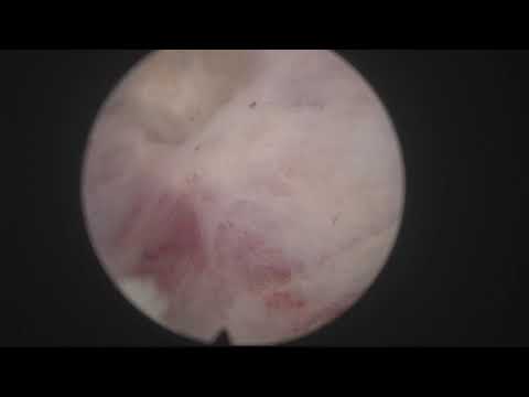
A MoLEP reoperation after TURP, in a patient found to have a stone within a bladder diverticulum. The published plan included removal of the stone, enucleation of residual benign prostatic tissue, biopsy of the diverticular wall and endoscopic treatment of the diverticulum. The description does not supply the biopsy result or long-term outcome.
Documented details and sources
Only details explicitly stated in the sources. Missing information does not mean it did not occur.
- MOSES / MoLEP
Real life MoLEP case 23: Redo after TURP with stone in diverticulum
- Redo surgery
Real life MoLEP case 23: Redo after TURP with stone in diverticulum
- After TURP
Real life MoLEP case 23: Redo after TURP with stone in diverticulum
- Bladder / diverticular stones
Real life MoLEP case 23: Redo after TURP with stone in diverticulum
- Bladder diverticulum
Real life MoLEP case 23: Redo after TURP with stone in diverticulum
Source checked: 2026-10-06.
Available transcript · English
Automatic YouTube captions, not clinically reviewed. Names, technical terms and numbers may contain errors.
They may contain transcription or translation errors; check the explanation in the video.
- hello again this is fernando estancia i have been out of the video production business for some time mainly because my recorder at the operating room fell to the floor and i was not able to record the narration of the operation while i was doing the operation and having to record this at home it's a little bit more time consuming so this is uh but i'm back this is the interesting case of a patient who was submitted to a trp uh in the past and he developed a stone in a bladder diverticulum so we discussed his options his voting was not too bad he was not severely bothered by by his symptoms but i told him that these stones inside the diverticulums can cause chronic inflammation of the bladder and
- that can lead to bladder tumors somehow so i didn't feel happy to leave that there and i we discussed that if he was going to go in the operating room again it was probably a good idea to finish the job after the the turp and so i started to use this moses fiber to treat the stone i was using one joule and 40 hertz which is the setting that i usually use for coagulation and the pedal and to my surprise there was a biological matrix that probably led to the formation of the course of the of the stone sorry [Music] and as we uh peeled off the external shell of of of the stone we could see that there was some biological stuff in the middle so probably a trp chip left behind at the time of the original trp
- operation so i guess yeah it is imperative when you when you do endoscopic surgery especially trp to check inside the diverticulums to see if there's any tissue left behind because this could be the consequence of uh forgetting you know a piece of tissue uh inside this this diverticulum so i thought it wasn't quite interesting case to show not only because of the stone and because of the course of the stone but also because i love these redo cases i think they're very interesting and also i think it's amazing how we can treat these cases perfectly with the in-block approach there you are so this was the fragmented stone most fragments still in the diverticulum so i like to evacuate fragments
- by gravity you know i feel the bladder i point to the stones i close the inflow and then i take out the internal sheath you see so the stones tend to be sucked or pushed out by the water coming out of the diverticulum or coming out of the bladder some of the stones went into the bladder but again they leave some time to feel the bladder and then i like to take them out with gravity this way it's quite um i think i find it nicer than using the the helical equator or the syringe because when you do the elec evacuator thing you the position of the scope is not known you don't know where it is you don't know where um [Music] if you're going to be sucking the bladder you know so there were some other small
- diverticulums and also i told the patient that we could try to do an endoscopic treatment of the diverticulum this is uh possibility for smaller diverticulums so that's the sphincter you can see there is residual tissue in the fossa some areas where let's say resected better than others apparently some there's a favorite side for surgeons you know like remember when we did trp uh you would feel more comfortable resecting in one side than the other side so maybe the job was better in one side than the other side so here you can see the marking of the of the white line and this is using moses and you know moses is a pulse modulation emission mode and the laser emits two pulses instead of one so this
- enhances the cutting properties and the coagulation properties so here you see the thing here is you have to respect the sphincter but you have to make sure that you remove everything so here i'm just trying to find out what the the white line should look like to try to do the the most perfect possible job you see trying to dissect the plane was not easy and this is mechanically i mean on both sides you see usually when you push in that region the plane opens for you but we mustn't forget that this man he is a re-operated man so most likely after trp the remaining prostate and the remaining adenoma probably undergo fibrosis and inflammation and [Music] it is not unusual to find that these redo cases are a little bit a little bit
- difficult because of that so this is one of the advantages of pulse modulation i think that the enhanced scanning properties of this laser uh help you know in the dissection of of these planes so here i was carefully judging you know what what is the proper plane we couldn't find i couldn't find a beautiful plane us would normally find and [Music] this is probably to do as well to the previous turp i guess when trp is performed many glands conduits are ducts sorry are uh obstructed i think and many of these residual glands are dilated and you will see that the quality of the plane is not very normal no like a virgin prostate that was never operated upon so here i am trying to progress a little
- bit laterally as well and trying to complete the the white land here i didn't mark the anterior part initially i thought it would make sense to check as i go you see this this is the marking i made the other side is not complete yet but you see how the adenoma is touching here i mean in some areas there's going to be less remaining tissue is going to be a thinner slice of remaining bph tissue or maybe in some areas you could be at the level of the interface between the capsule and the abnormal so that's what makes this redo cases a little bit more challenging and more interesting so here as you can see coming up carefully of course i want to protect the sphincter the completely and to be sure that patient
- will have a very good continence and here coming up towards 12 and trying to take the working uh area a little bit further inside you know so we're trying to get away from the sphincter to to respect it to stay close to the sphincter the least possible time so here again coming up coming up coming up towards 12 o'clock this was the um area that was uh best uh resected probably i was checking the water or something when i was stopping like that so i took out my eyes from from the screen and do something often you you have to do that and uh here as you see we're coming up from both sides trying to connect at 12 o'clock and to try to develop an anterior plane you see this is the anterior plane now coming down
- very difficult uh differentiation of the plane so i'm trying to use the energy to let the energy decide where uh the plane is going to go you know holmium laser has a very high peak power that produces this this uh cavitation bubble in front of the fiber that produces a shock wave that is going to dilate or distend or dissect sorry the the plane between adenoma and capsule but here having had a previous drp there is a lot of fibrosis still you can see that the energy is opening up the plane just carefully you know checking that it's the proper plane or very near to it [Music] also another very nice aspect of pulse modulation is the nice first pass hemistasis which allows to do a very nice operation
- you can see here the tissue quality is very strange the capsule quality is different than the unusual and [Music] many times we find these dilated spaces probably obstructed glands after the epithelialization of the after trp so now of course the sphincter is totally safe i'm just going around trying to follow the plane to see if we can approach the bladder neck progressively circumferentially you see going around the whole anomaly try to follow the plane yeah that looks good but carefully yeah because it is important when you do hollap to progress and to be able to detect very early if you're going too deep in your dissection especially when the plane is altered like here due to this uh you know previous inflammation sometimes
- in these cases you can dig a little bit deep in the peripheral zone posteriorly so you have to be careful and and progress and if you detect that you're going too deep then correct before it's uh too late and that's why you have to be careful keep the fiber moving dynamically so you don't stay too long in the same spot because if you stay too long in the same spot you might deepen too much so keep moving keep moving keep trying to do superficial improvements of the dissection and don't ah here here's the access to the bladder anteriorly and now we have to check we have to check what's going on here brilliant that is anterior that's the other side coming up you know it's always the same a nucleation
- and blocking nucleation makes the procedure very very easy and very safe for the for the sphincter and it becomes very very familiar and very intuitive as well because you are just going around the the noma as if you were peeling an orange of course nearing the bladder neck we find more fibrosis you see tissues are hard difficult to cut and but slowly we can progress and the objective of this operation was to try to make sure that this man [Music] who was operated once and now has to be operated a second time will never need a third time if it's possible so trying to remove the residual and normative tissue that will certainly grow if it is left there maybe um causing the need for a third operation
- in the future if we only removed the stone so here that is the uo so we are quite far you can see that the surface is a little bit rough it's not as uniform as we are used to see but as i say this is possibly due to the post-operative inflammation after trp and later fibrotic processes and that's why we see these dilated glands and sticky parts where developing the plane is a little bit more difficult so yeah you always have to think you know here i thought maybe i'm leaving this amount of tissue i didn't catch the good plane and yeah it proved you see that i could find a better plane so you have to challenge your own plane sometimes in a situation like that you might find that you're going a little bit deep so
- then you have to stop and uh follow the previous path but this is the way you need to challenge the try to find what is the best the best plane because you don't want to leave significant residual bph tissue in in the fossa you see how maybe in some areas you can go near the peripheral zone laterally and if you see that you're going too deep then you have to correct a little bit and medialize the fiber the incision so the targeting of the laser has to to be corrected to try to adapt to the anatomy so this is uh now coming up to try to connect with the already opened bladder neck here we find typically this area which is already a little bit difficult uh to detach in in a nucleation the the six o'clock area
- you know between adenoma and bladder neck many times there's a steep curve you know a change in the direction of the plant as you approach the bladder neck and the bladder neck six o'clock is more like a wall so there's a hard transition from the floor of the prostate and then you approach this wall and then it's not impossible here you can see a little bit of fat so it's not difficult to to cut through the wall and and get into the the retrotrigonal space so you have to be very very careful maybe take it from medial to sorry from the lateral aspect towards the medial trying to keep in very very almost in contact with the normal fire very close to the enormous so that you can detach it but not penetrate deep in this
- in this region this is all complicated in trp cases because usually the middle low area is the best let's say resected and the deepest resection area and it can be sticky sometimes so that's again coming up from the side trying to connect to the lateral dissection trying to go from side to side trying to progress it's a matter of improving the situation every minute it's a matter of patience you know you shouldn't hurry just take it easy progress slowly this would this will take you uh to the fastest possible result i think if you are careful and you progress without compromising the procedure and take your time to cut and to dissect carefully also if you see as we have seen a little bit of fat in a
- small area don't panic just keep working correct the plane usually it has no consequence to go a little bit deep in a region of the capsule it's important to stop in time so you don't do a massive perforation in which case probably it's better to put a catheter and come back another day but here we are progressing yeah now the norma's tilted into the bladder and there is the the bladder neck that we have to cut very fibrous tissue but nearing the completion of the nucleation of the residual tissue after trp so here you can see how the bladder neck is completed and the adenoma is pushed into the bladder that's a sphincter we have managed to preserve the sphincter beautifully and there is some minor
- residual bleeding that's a small nodule so i'm going to vaporize it see just below the nodule it looks a little bit flimsy so you can spend some moments to make sure that you vaporize these small nodules like that some vessels going not too much the next thing i'm going to do is i'm going to try to get in with a biopsy device to take biopsies from the mucosa of the diverticulum we want to see if these changes we have seen in the diverticulum and that's a norma you can see that it's a doughnut it's a circumferential piece so we have managed to do an unblock a nucleation of the residual tissue after turp this is an unedited video so i'm not cutting anything just showing you the real time real-time work and here
- i changed i think no and the next thing of course we're going to do is the musculation of the piece so here i changed to the musculature i was trying to say that the plan was to take a biopsy of the of the diverticulum i tried to focus for a moment and often this post trp tissue is also very fibrous and sometimes difficult to morsel it there's a lot of fibrosis it's uh tissue with the consistence of leather you know it very very difficult to cut but in this case it was not so bad just took a little bit of uh time to mercilate just have to be careful to keep the bladder full and to keep the blade away from the from the bladder wall but uh this is the perennial oscillator i love it i think it's
- excellent really fast and it really really helps to have a good more slater because it shortens the operation time significantly here you can see that we did the stone in the diverticulum and the nucleation it's been now 24 minutes so these operations are not lengthy anymore because mainly mainly because of the most relation but i have to say that after using pulse modulation we have noticed that we are faster because we don't spend too much time in hemostatic maneuvers during a nucleation so we can just go go go go dissect dissect dissect and euclid and then we'll have to spend a lot of time doing additional coagulation so this is the end of the adenomatous tissue now coming out typically they most later choose the
- easy parts first and the fibrous parts are more resistant so they're left for the end and this is when we have these beach balls that are a little bit more difficult to to more slate but in this case it was perfectly possible that is one of the diverticulums for a moment i thought would it be possible to suck the diverticulum inside the mucosa inside the bladder and then mostly just a crazy thought i thought that's better to not do that and then of course we changed here you will see there's a little bit of bleeding and that's because we decompressed the bladder completely and of course i didn't spend probably enough time to do perfect hemostasis but i thought i'm going to come in with our scope so there you see
- there's some accumulation of blood here i think i was going to try to to take the biopsy from the diverticulums just found the diverticulum look at this changes you see what is that so i'm sorry for my voice here i think i was trying to put the that's the bladder neck where are you diverticulum for a moment i couldn't find it so i took out my my view from the diverticulum and then i had to put the biopsy forceps called biopsy forceps and there it is look at that ah stones inside dirty articulums are not good news so i decided to take some biopsy to send to the pathologist and then of course the other interesting point of this video is that i i'm going to try to do the endoscopic treatment of the diverticulum
- you can try to treat this mid-sized diverticulums endoscopically and how is that accomplished well you have to go back to the old receptor scope and i thought this was third sample and as i said we're going to change it now for a resectoscope see there's a light hematuria usually if you put a catheter that completely stops and it's not very relevant but as i was going to go in with our sector's monopolar situscope i thought there's no there's no need to be very very thorough with with the hemostasis and we can finalize with with the recycle scope you see also of course this tissue probably is more inflammatory than unusual and more prone to bleeding a little bit so i thought let's spend two minutes
- perfecting hemistasis in the fossa and then i will go to the diverticulum many times i think this these bleeders for some reason when we put a catheter and the prosthetic capsule collapses they tend to stop and it is the distension of the fossa that keeps them bleeding that's the venus venus bleeding if there are small arteries of course it's a different matter but yeah nothing nothing serious so i just wanted to have a little bit better visibility in the bladder that was clot that was already forming this bladder neck okay so now where are you again here so this is the interesting part i think of this video and this is the endoscopic treatment of a diverticulum hoping that this is only inflammation
- i'm going to vulgarate the whole mucosa of the diverticulum and systematically make sure that all the mucosa inside the diverticulum is vulgarated coagulated um for some reason i didn't think about doing this with the laser itself but which probably it's a possibility just photocoagulate rather than electrocoagulate but here i was [Music] trying to remove the inflamed mucosa and the idea is that if you coagulate the whole diverticulum like that and you keep a catheter for three or four days connected to a bag so the bladder is empty this sticky surface is going to remain collapsed and possibly it will connect again this perivesical tissue will connect again with the bladder wall and when re-epithelization
- takes place again the size of the diverticulum will be less than the original this is the endoscopic treatment of bladder diverticulums so here i'm trying to see that all the wall of the diverticulum is vulgarated and if if i if i have a chance to scope the patient in some other moment i think i will try to add or connect some further information to this video to see if this really works but you see now with the mucosa totally gone and the bladder totally empty it's very likely that we will now there were some stones there so i did the same maneuver again just removing you see the inner sheath closing the inflow and letting the gravity or the outflow water from the from the bladder uh take out this this remaining stones
- searching for other diverticulums this is the one i already treated there's a small one there that i lost this is another one so i'm going to try to do the same here see if we reduce this diverticulums and i hope it works i think it's interesting to see how to torp that is considered by many as the best endoscopic treatment for bph also has its problems and in this case this man has a stone in a diverticulum uh due to a forgotten chip of tissue inside the diverticulum stone has formed and an inflammatory or maybe these plastic changes have taken place in the mucosa of the diverticulum so this is my treatment for this case two diverticulums are vulgarated coagulated and we will keep a catheter for three
- days four days to see if we can end with this or improve the situation i hope you enjoyed the videos and thank you very much for your attention once again you