
Surgery
Real-life HoLEP 21: Diverticular bladder with stones
Dr. Fernando Gómez Sancha · ICUA
Dr. Fernando Gómez Sancha · YouTube
Language: English. YouTube audio metadata.
Surgical content for professionals. Contains footage of an operation.
Playback connects to YouTube. Your search query is not sent to the player.
About this video
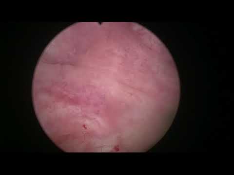
An en-bloc HoLEP in a patient with urinary retention and a documented residual of two litres. The description also identifies a diverticular bladder with stones. It does not describe the subsequent management of each diverticulum or stone.
Documented details and sources
Only details explicitly stated in the sources. Missing information does not mean it did not occur.
- En bloc
We decided to do an En-bloc HoLEP. He had a diverticular bladder with stones, and this is the video.
- Urinary retention
This man had a retention of urine with a 2 litre residual. We decided to do an En-bloc HoLEP. He had a diverticular bladder with stones, and this is the video.
- Bladder / diverticular stones
Real-life HoLEP 21: Diverticular bladder with stones
- Bladder diverticulum
Real-life HoLEP 21: Diverticular bladder with stones
Source checked: 2026-10-06.
Available transcript · English
Automatic YouTube captions, not clinically reviewed. Names, technical terms and numbers may contain errors. Check them against the video; complete audio coverage has not been verified.
Selected spellings of enucleation, morcellation and holmium terms have been edited. This is not a full audio or clinical review; the original captions remain the source.
They may contain transcription or translation errors; check the explanation in the video.
- hi this is fernando romer sancha and this case we recorded it in the operating room and i thought i was also recording the audio but i forgot to turn it on so for the duration of the operation i was commenting on it but it couldn't be recorded so i but it's rather interesting case and i i thought it was worth uploading it to my youtube channel and so i decided to to to to do a voice over of the video this patient is an 84 year old man he had a retention of urine with a two liter residual and was wearing a catheter they couldn't
- remove the catheter and he came to see me he is a relatively high risk patient and i told him despite your bladder diverticulums we're going to do a prostate enucleation and we will see how you do if the diverticulums do not pose a problem a significant problem and you can empty reasonably and have a reasonable quality of life after the operation maybe we don't need to treat the directions let's see that would entail a little bit more aggressive surgery so we we did this as a first step you can see that i'm using
- the outflow of water so i fill up the diverticulum with water and then i use the outflow to carry the stones outside luckily these stone fragments were relatively small and they could come out so gradually i would fill in the diverticulum and then let it empty and bring the stones out it's interesting to see the inner sheath of the scope you see it's a metallic tip that i use and i love and it's a little bit damaged by the laser um fiber and this happens when by mistake you fire the laser with the
- with the fiber inside the scope luckily this metallic um tip can take it it is much worse there's another tip which is ceramic and this ceramic tip can easily be broken so when i was doing green light and nucleation i used to do mechanical dissection of the plane with the tip of the scope once i was invited to do uh an operation uh somewhere and when i got to the operating room they had already prepared the patient and the endoscope was inside already so
- i couldn't have a look at the tip of the scope but it was a ceramic tip and it was broken by laser by laser blasts so instead of having a smooth surface it was quite damaged it was like a sole you know with teeth and when i started doing the mechanical dissection i thought what's going on with this plane you know it bleeds all the time i cannot produce a nice looking plane and the thing is that i was using a tip that was uh severely damaged and that's why i always have a look at my tip before i start the operation because the
- patient had a little bit of a rough time because of the bleeding and afterwards and um and i realized that if you're going to do mechanical enucleation the tip of the scope has to be totally smooth and without any possibility of damaging the plane so that this is one of the lessons you learn painfully and so um since then i like much more this metallic tip in this case i changed the working element of the laser for the resictorscope loop for a moment to extract the the last stones that were a little bit bigger and then
- trying to complete the instruction of the of the fragments if if there are minimal fragments probably you know they could be passed after the operation but in a man with a diverticulum i think or with many diverticulums i think it's better to be thorough and try to remove uh all the stone fragments before starting the indication it took a little while you know it's about six minutes now of uh surgery but it's almost done i think that fragment finally wanted to come out it was small enough that the loop couldn't grab it properly
- all right you see it's a heavily you know traviculated bladder and with multiple small diverticulums and not so small maybe a lot of edema due to the catheter here i was trying to check that there was no more fragments and the rest of the diverticulums
- and yeah those are the last tiny bits of stone coming out before the the nucleation starts so i think again a last look again is a receptoscope loop we always have a receptor scope on the table for usually for hemostasis but it can be useful in situations like this or even at the end of the operation if there's a residual fragment sometimes the loop is good because you can catch it and extract it relatively fast so the receptor scope keeps being useful but maybe not not so much for turp but for auxiliary maneuvers and
- to complement what we're doing with the lasers so that's the laser fiber again and the size of the prostate was about 80 grams i think and again we start with the usual strategy to mark the lower aspect sometimes in patients who have been catheterized it is not so easy to tell where's the sphincter because after wearing the catheter the sphincter and the apical image sometimes it's not so easy to to to distinguish so maybe that's why i was a little bit conservative here but that's this thing edge clearly there and that's uh
- trying to to mark the white line now you see this uh prosthetic urethra is quite inflammatory because of the catheter possibly and we had a lot of mucosal bleeders and this can be a nuisance at the beginning of the operation sometimes but i try to complete the white line complete the white line deepen it a little bit if it's possible here again i think it's very important to cut this attachments of the sphincter with the apex in the posterior aspect at the apex that's a small dissecting maneuver you see when you do this kind of mechanical
- dissection as i said you have to make sure that the tip of your instrument is smooth because if it's pointy if it has irregularities then you could do more more damage than than you would want to do so that's a little bit of laser development of the plane we know that we are in the good plane but just developing a little bit further and again on the other side the same maneuver all right cutting the fibers that are joining the apex posteriorly to the sphincter and and then entering the plane with a little bit of mechanical dissection very smooth pushing and then of course the development of the plane
- if if you do mechanical dissection then you'll have to do hemostasis later if you do laser dissection then the hemostasis tends to happen while you are dissecting so that's why i have limited um the mechanical dissection maneuvers but of course sometimes they become very handy and very nice to determine where the plane is alright so once there's a single plane posteriorly and a small cavity i usually put the fiber 12 o'clock which is my favorite position and then i start developing the posterior line of attack so this line of attack or this line of dissection is extremely helpful to
- to know where is the plane to orient yourself you see that instead of deepening the dissection in one point i like to do very wide very wide movements from side to side to keep a very very wide line of attack that helps determining where is the good plane in this case the plane develops very nicely and it's very easy to see how the adonoma is detaching from the capsule you can see that the fiber the laser fiber is trembling a little bit and this is because i don't use any support for the fiber i don't use the the cons [Music]
- device you know the trigger that allows you to use the fiber as if it was a resectoscope loop i have my free hand holding the the fiber and this allows me to have a very very very sensitive control of the tip of the fiber i can push it in or bring it out with the extreme precision because i'm using my fingertips to to hold it and uh it gives me extraordinary freedom you know i went to a course where i had to do i had to do an operation using the the const device and i felt severely impaired to to to carry out this technique because it was somewhat limiting at the end of
- course i managed but i i love this freehand manipulation of the fiber and there is some trembling as you can see but it's not i would say clinically meaningful i think this tremor of the fiber doesn't limit my ability to to to [Music] to perform the operation all right so this is the ascension towards one of the sides you see so it's very nice to to follow the good plane all the way up but the same way we cut the posterior fibers joining the sphincter to the apex you have to do the same anteriorly
- so this is already quite uh well advanced that's a 12 o'clock region you see all around the sphincter and following the white line one has to deepen the incision a little bit first and then once you have cut three or four millimeters in depth then you have to start thinking and searching for the correct plane this is not going to leave any significant amount of tissue attached to the sphincter but it will make sure that the sphincter is left behind it's left distally you see here cutting these fibers at 12 o'clock on the other side and i don't mind of the plane there a
- little just when we are a little bit more inside then i will worry about the the right plane so this early apical dissection uh it's a very protective of the external sphincter you see this is the is the white line on the other side that's the good plane you see i'm not now concentrating on the good plane i'm concentrating on the touching the apex from the sphincter so i will do this incision in the edge of the sphincter onto the adenoma you see on the adenoma and then once the axis has been established when once we have been able to leave the sphincter a little bit behind then i will search for the good plane and
- this ascending dissection will aim to connect the plane to the other side to the dissection we did on the other side it's always very careful always having the sphincter in consideration we want to do a very delicate access to the to the to the plane but respecting the sphincter and especially as uh respecting sphincters mucosa because i feel that [Music] ripping the mucosa off the sphincter can cause temporary incontinence that has been reported very often in papers on holleb and the truth is
- that we have a extremely low incidence of stress incontinence you know we don't really anticipate that incontinence is going to happen and we don't tell the patients they have to do careful exercises or anything like that because in our everyday practice what we see is that patients do not have stress incontinence after this operation only very very exceptionally and when when we have seen patients who tell you i have incontinence uh after this operation it's usually due to a hyperactive bladder patients who
- already were incontinent before the operation because they had this terrible urge to void and to go to the toilet [Music] these patients can have the same symptoms for a while during the operation but we don't expect to have a stress incontinence because the protection of the sun sphincter we do is is excellent so that's you see the anterior plane the connection has already been established from side to side you see the the anterior plane is being developed and the line of attack is now circumferential around the adenoma so it is possible to to follow this line of attack you see that the plane was very beautiful in the lower aspects but maybe not so beautiful in the lateral
- [Music] aspect so it's very helpful to have this line of attack because it orients orients the the dissection very nicely of course you have to know that uh the plane is going to be circumferential so instead of doing straight lines we're going to follow a curved dissection curved line trying to adapt to the to the shape of the prostate so you can see how the camera is or the scope is always trying to keep the line of attack in the middle of the screen and this gives us the vision of the capsule on one side on the noma on the other side and
- one has to concentrate in trying to keep in the good plane sometimes it's very obvious that the plane is good because the dissection is very easy it's very natural and uh the characteristics of the plane are obvious now and you can see that you are in the right in the right plane and in the right depth of dissection sometimes it's not so obvious and then you have to well you have to do your best and it's a very [Music] it's a very good help to have this circumferential line of attack because if the plane is good let's say two centimeters on the right
- or on the left or up or down we cannot be very far far from the good plane so i concentrate on the details you know on the information that i can get from what i'm seeing you know the characteristics of the capsule uh the color you know the capsule has a white whiter color than the adnoma i think if you see yellow tissue on the capsule that's usually adenoma and with this information that we can get from watching the characteristics of the plane you can progress in the operation trying to keep
- in this interface between adenoma and capsule i think maybe if you have watched uh other videos this is uh quite repetitive but uh this is the principle of of of the separation here we are reaching the bladder neck we're reaching the bladder neck and we are carefully carefully trying to find the way to enter the bladder between the abnormal and the capsule so by following the plane from the sides coming up see it looks a little bit flimsy here the danger could be
- to catch the capsule anteriorly and exits not inside the bladder but exit anterior to the ladder so it's also important to acknowledge that we are dissecting a pseudospherical anomalous tissue and not only the line of attack is circumferential but also we have to be conscious of the 3d shape of the prostate so when we are approaching the bladder neck we have to correct the angle of dissection and when we are doing the dissection
- anteriorly of course the plane is going to descend towards the bladder neck in the posterior aspect where we are dissecting now the plane is going to start ascending towards the bladder neck so the idea is that as we get closer to the bladder neck the fiber has to get progressively closer to the adenoma to correct the angle of dissection you see here if you fire against the line of attack itself there's a risk that you could go inside the capsule that's why i try to keep the fiber
- closer to the edge of the adnoma closer to the edge of the adnoma and not not so much firing towards the line of attack itself as i did at the beginning because the angle of the dissection is changing you see we are ascending now the posterior capsule is changing direction now it's ascending towards the bladder neck so as we progress it's no longer descending as it does at the beginning so these are to me the crucial points of technique of this operation you know you have to know that if you want to dissect the plane and not violate the capsule you have to keep the fiber closer to the abnormal closer to the adenoma you see always very close to the adenoma if you
- dissect a little then you have to correct and go again close to the abnorma to to make sure that the energy is only coagulating the capsule but not damaging it you see if you get too close to the capsule and if you fire the laser there maybe you can penetrate into the capsule small penetrations into the capsule are not dangerous and not a problem but if they are bigger then there could be trouble this is hemostasis all the time you spend doing hemostasis is well spent i think especially if you do your hemostasis as you progress with your dissection this is again the lateral plane coming
- coming closer to the bladder neck area you see the waters now irrigating this space between the capsule and the enormous but it's not it's not entering the bladder because that's why we get so good visibility you know one of the trouble people had with with hollop was bad visibility during the operation and this technique the unblock technique really improves this this visibility because we are irrigating a very small space the space that we are developing between adenoma and capsule here we are getting closer to the bladder neck but still
- it is not very clear you see sometimes things look a little bit strange and you're not sure where is the entry to the bladder so one thing you can do is you can go towards 12 o'clock and make a 12 o'clock incision there no need to do it do a complete incision but just a little bit at the bladder neck and many times you will connect the space you will connect with the anterior space here i didn't really connect but just going inside the urethra gave me the information that bladder neck was probably around there and there it was all right so some people are afraid of this step
- you know entering the bladder uh without any you know clear reference i always say that the direction of the fibers changes there from circular fibers to vertical fibers but well this is a nice tip if you encounter trouble getting in if you're not sure it's always prudent to go inside the prosthetic urethra and make a 12 o'clock incision too to connect a little bit irregular surface but that is now the the bladder neck and we have to cut the bladder neck trying to follow its circumferential shape
- you see that my fiber is always at 12 o'clock i i don't like to rotate the scope i think rotating the fiber is a little bit confusing many many people do and that's okay i i think everyone has to find their favorite way but i think keeping the fire at 12 o'clock simplifies a lot the handling of the scope and the fiber so you don't need to rotate you just need to focus on where you are positioning the the scope and the fiber and also i think when you rotate the scope sometimes the camera gets a little bit off and here for example i know exactly
- where i am this is 12 o'clock and uh because the camera is telling me the truth all the time all right so that's why i choose to use this at 12 o'clock and it's uh absolutely possible to [Music] do the circumferential dissection with the five or 12 o'clock i don't see there's any really significant advantage to to rotate the scope but as i said this is a personal preference so that's the lateral plane on the other side and trying to connect this lateral plane to the bladder neck incision we just made ladder neck opening once the bladder neck is opened the
- irrigation of this small space that i was referring to is no longer the same in the sense that there is now irrigation fluid going in the bladder and coming out the bladder but also if there's bleeding there will be blood going into the bladder and maybe it's not so easy to to take it out to wash it out as it was before when we were irrigating a closed small space there there's the bladder neck again it's coming up coming up towards the bladder neck you know after sometimes this this this operation becomes a piece of cake you know
- i think it has a learning curve i think you have to learn it you have to get used to the scope and the fiber you have to get used to the anatomy which is totally different from what we have seen with trp because the trp had a totally different uh visualization of prosthetic anatomy and i was remembering peter gilling in one congress saying to someone you have never seen the capsule of the prostate i think that was a little bit radical but of course the anatomy is a little bit different and you have to
- to learn how how to interpret this this anatomy but and if if you see a lot of operations i think if you see a lot of videos like this i think you will get more and more knowledgeable and you will be better equipped to to do your operations i find that navigating this circumferential plane is something you have to learn and you have to get better at and that's nearly the end of the nucleation you can see it's a very fast technique the sphincter was dissected at the beginning it was respected completely and also a fast technique i think is a good thing for the urethra it's a good thing for sphincter
- you know being able to complete these operations in in such a short time it's been 31 minutes of surgery up to now and you can see the mucosa of the sphincter has been preserved and the white line that we marked has been absolutely the the landmark that we have respected uh during the operation so that's a little bit of hemostasis i was using the 100 watt console the old console and so if you want to change the settings you have to stop and you have to so i often try to coagulate with the same settings as i used to to do the the nucleation and that means that you have to separate the fiber a
- little bit more from the tissue to get the coagulative effect this is the final detachment of the anoma so that's a uh from one side the uo from the other side and a little bit final hemistasis so the purpose of uploading these videos is to try to give those of you who are learning a chance to see many procedures to see how different the anatomy looks from patient to patient the quality of the plane is not always the same and how you have to adapt to that i think the the unblock technique is is uh
- excellent it's probably easier to learn you know when i teach people i i try to do the epic liberation first and then leave them to do the rest of the dissection because if you think about it the sphincter is already safe and you can do some learning you know residents and people who are learning can work relatively safely around the ednama you know getting used to manipulating the scope in a pretty safe let's say training model huh of course as i said you have to see a lot of videos you have to understand the anatomy you have to do your cases i mean you have to see someone doing the procedure i mean it's always better to see the live operation and by the surgeon than to see a video
- but that's some sometimes not practical or not possible and then of course in your first cases you must have a mentor who helps you for example initial cases someone does the the epic liberation and then you do the rest and as you get more confident then it's not such a difficult thing to do to to liberate the apex but of course it needs some getting used to and the manipulation of the scope is a little bit different from how you manipulate the scope in during trp so this is now the change of instrument uh to start the morcellation you see there's quite good visibility inside the bladder a little bit of bloody um medium so what i like to do is to check
- where's the bladder neck and then i would elevate the tip of the the blade a little bit to try to keep in the center of the the bladder neck and then i like this situation where i can see the blade i can see the adenoma on top and i can see these two black triangles on the side of the blade that tell me that the bladder wall is far away with these fast most laters this is a perennial system we can do a very fast more solution we measured 11 grams of tissue per minute so if this prostate had an estimation of 80 grams maybe the adenoma was 60 probably in five or six minutes the more slation will be totally
- finished so it has radically changed this operation and if you think about it an 80 gram prostate with stones in the diverticulum will take around 40 minutes to complete so it's very very competitive with trp and i cannot find any reason why trp would be better than this for example in this patient has been almost no bleeding we have seen the anatomy we have removed the whole adnoma and [Music] so i understand it is difficult to change when you're used to doing something when you're used to hearing that this is the gold standard and
- but i i would like to encourage you if you if you don't do hollap to to try it you know if if you don't have a laser you have to i don't know steal it to get one you know do whatever it has to be done so you can start doing this immediately because these are patients that are super happy we will remove the catheter the next morning in every patient apparently now with the new pulse modulation hollop devices it is possible to have much better hemostasis and remove the catheter earlier or even [Music] you know not need irrigation which means that once the patient is ambulant he can leave
- the hospital with a catheter go home and then come the next day to have it removed in the clinic so this is opening the path to ambulatory surgery uh even with a nucleation for us it's a little bit difficult because we work in a private hospital so private patients do not like to be pushed home you know with a catheter and [Music] it's maybe a little bit different than public health systems where the patient is a little bit less demanding and more he has to obey what they tell him more than in a private environment where the patient has this client let's say consideration no we have to look after our patients
- and sending them them home with a catheter may be is difficult to explain when they can stay overnight and then have the catheter rooms removed the day after and here is more or less the conclusion of the case i think there's very little abnormal left to to morcellate and that is the fossa very nice hemostasis i hope you enjoyed the case