
Surgery
Real Life MoLEP case number 14: Patient in retention with Gleason 6 prostate cancer
Dr. Fernando Gómez Sancha · ICUA
Dr. Fernando Gómez Sancha · YouTube
Language: English. YouTube audio metadata.
Surgical content for professionals. Contains footage of an operation.
Playback connects to YouTube. Your search query is not sent to the player.
About this video
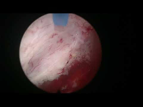
An en-bloc MOSES HoLEP in a patient with refractory urinary retention after treatment of a stone in a left ureterocele. The patient was under active surveillance for prostate cancer described as Gleason 6 in the title and ISUP grade I in the description. The published account identifies a three-lobed prostate and the shared decision to perform enucleation; it does not describe HoLEP as treatment of the cancer.
Documented details and sources
Only details explicitly stated in the sources. Missing information does not mean it did not occur.
- En bloc
We decided together to perform an en bloc enucleation. Nice MOSES HoLEP case in a man with a three-lobed prostate.
- MOSES / MoLEP
Real Life MoLEP case number 14: Patient in retention with Gleason 6 prostate cancer
- Urinary retention
Real Life MoLEP case number 14: Patient in retention with Gleason 6 prostate cancer
- Coexisting prostate cancer
Real Life MoLEP case number 14: Patient in retention with Gleason 6 prostate cancer
Source checked: 2026-10-06.
Available transcript · English
Subtitles exported from the original-language YouTube Studio editor. Not clinically reviewed; source identity and timestamps verified, full audio coverage not verified.
Selected spellings of enucleation, morcellation and holmium terms have been edited. This is not a full audio or clinical review; the original captions remain the source.
They may contain transcription or translation errors; check the explanation in the video.
- okay so this is the last case of today i think the access is a little bit tight but no it's good oh yeah we have big urethra in spain maybe so this is the sphincter h more or less and you can tell this was supposed to be a very large gland maybe it's very wide ah it's not too long yours are close to the edge so we have to be careful later on bladder a little bit trabeculated no but this is a very standard very common in the patients we operate snow so let's put the fiber in that's the barrier again and here you can see the edge of the sphincter now here i'm going to mark here the the white line huh so i'm going to try to develop this this white line there's some mucosal bleeding it's quite common
- so sometimes you can even have trouble let's say finishing the the landmark the marking of the of the white line there's a little there's a lot of lot like now where you know it comes to a point where you don't see very well my advice is to go in the plane now to look for the plane and start a nucleating if uh on the contrary you can you can see very well my advice is to develop let's say a groove you see following the white line if you deepen the white line a little bit you know you can you can develop a groove that establishes a separation between the abnormal and the apex let's go into the plane so this is a very montanan here you see if you if you push a little bit with your scope you're going to enter the proper plane
- very easily let me cut and coagulate here i want to release the the attachments of the sphincter to the adnoma from six o'clock to to nine o'clock in this side and then on the other side i'd like to do the same it's following the the white line the typical maneuver to to enter the the proper plane is to put the scope very close in close proximity to the very montana and then push a little bit sideways uh lateral but you can you can do a little bit of mechanical dissection on the apex that will give you the clue of uh where is the where is the where is the good plane of course when you have localized the plane in both sides it's only logical to try to connect them in the midline so you cut on top of the very montanum
- the beginning of the operation sometimes you can have bad visibility but this will very soon change as you will see of course you can try to spend a little time trying to find out where is the bleeding coming from and uh try to do some hemostasis but most of the times you can just progress and hemostasis will take place also a lot of the bleeding is coming from the adenoma and as you would say detach the adenoma from the capsule you are sectioning the vessels that yeah so that's the posterior plane i'm going to put the fiber at 12 o'clock and i'm going to follow the nice section a little bit further of course if the bleeding continues i will try to do some hemostasis before progressing here you can see that i'm developing the posterior plane trying to split the screen
- into trying to keep the line of attack the line of dissection in the middle of the screen and at the beginning of the procedure my let's say aim is going to be to lace against the line here for example you see the capsule is getting a little bit thin here we will have to get let's say closer to the adenoma but i think this this is probably enough posterior dissection for the moment see where the bleeding is coming from let's see if we can stop it it's a little bit it's a little bit of a nuisance no it's a little bit epical optical bleeding okay there's a vessel there let's see if we can control that one there's a number of them huh okay so it's going to be a little bit bloody anyway this is 12 o'clock this is the white line
- we marked so the next and this is the mucosa of the sphincter you see so the next step is going to be to cut on the prostate to try to deepen let's say the the white line for three four millimeters five millimeters something like that i'm not trying to develop the plane just i'm trying to to cut yeah the adherences of the uh apex to the to the sphincter and then i'm going to try to look for the good plane huh this looked like a bit of tissue that was it's a little bit uncomfortable we don't have very good visibility for the moment it will get better now let's see if we can we can follow the the nice plane we get some better neurostasis well there we are trying to come up that looks like a normal
- trying to go from laterally around the apex here if you come out to see yeah yeah it's clear that we have to cut again a little bit on the prostate horizontally below the center to get to the let's say 12 o'clock fibers here huh this way we we leave the sphincter behind us now we can try to say conquer the peak of the arenoma at 12 o'clock progressively coming up coming up coming up but after having released the sphincter from the apex and the more most distal parts this is again a little bit of tissue on the apex coming towards 12 o'clock you see here is maybe in a case like this maybe with moses you would have the same initial bleeding but maybe the control of hemostasis would be a little bit better
- or easier no just doing the same work and doing the same steps you know so here we're coming up up up i try to look at the direction of the fibers here you see because that gives you the clue or where the plane is going sometimes we decide what to do based on tiny anatomical details you know that you have to be searching for and looking for of course you develop this understanding of the anatomy with some experience there we are so for the moment we released a big part of the noma coming towards 12 o'clock this is uh 12 o'clock fibers maybe we can complete the cut here the visibility is not so good okay the water is not uh particularly low so we have these apical bleeders okay so this is sphincter this is uh the white line we marked
- and again i'm going to cut on the tissue a little bit on the prostate here we are following a good a good a good cut and then now we look for the good plane now this is a good plane so we gain a lot of access by doing that incision on the prostate now here we are you see why i find it useful to develop the posterior plane a little bit because when you're doing this dissection of the lateral aspect you get orientation in the in the lower part in the posterior here we're coming up up up let's see what we have here you see this is it seems to be okay 12 o'clock although the visibility here is falling very fast so let's try to develop this lateral plane a little bit further this allows the tip of the adenoma to fall
- down you know it comes down you know my scope can go here we are lowering the tip you see so these fibers that sometimes are a little bit parallel to the to the laser fiber become verticalized you see because i'm pushing the nose or the tip of the of the adenoma down you see all these fibers become more accessible now we can cut them just because i positioned my scope on this on the lateral aspect we are trying to push down the adenoma and let's see if we can cross over towards the other side let's come out and check it yeah it looks like a great uh plane and now we have connected huh we finally released the apex completely we are in a good place we can continue our dissection now uh circumferentially
- there we are that's very good now we are connecting you see this is a normal let's say view of the capsule some people say that anteriorly there's no good plane but i disagree i think sometimes you find beautiful plane anteriorly as well you see i'm going to try to of course you have to find a speed of dissection that provides you nice developing on the of the plane but at the same time nice hemostasis you know if you rush it too much you can find that there are many bleeders going on at the same time and then it might be difficult to establish anostasis properly no it will take more time you will regret that you did the dissection very fast sometimes you get carried away with how good the plane is and how the dissection is progressing but then of course you leave
- too many vessels small vessels but too many at the same time you know so it's important to let's say carry a good hemistasis as you go and recheck it because many times there is some degree of spasm of the vessels you know when you cut through them and initially they don't bleed but then they start bleeding a little bit so of course we don't want to go too slow you know generating uh very very exaggerated hemostasis snow and uh widening of the whole fossa you know it's not usually necessary so you have to try to find the right balance now for you of speed of dissection quality of hemostasis but in general you see now we got excellent visibility now we have very good visibility and that's because we are irrigating a very small space
- you know just the space between the anomala and the capsule so that allows us to see very well and if there's any blood in the irrigation fluid will clear it you know just being a little bit careful with the hemostasis usually you can progress reasonably fast also you can see that i'm not lifting the pedal my foot from the pedal i am dissecting let's say continuously continuously all the time just very brief stops when i have to check something and i have to come back to check stuff but that makes this operation extremely fast you see i'm just progressing stopping to crackle it not too much not too little just uh enough and say in here you see the the fact that we are dissecting a circumferential plane
- makes it very easy to recognize i mean in some area you're not totally sure of maybe you see this and you think this is a good plane okay let's go up a little bit let's go down a little bit you see this is going to give you some orientation so of course if it becomes let's say if it looks deep or if you if it looks as if you're getting deeper and deeper you can correct you see by getting closer to the abnorma and then you leave you see a little bit of here is where we changed now we were following this plane it looked a little bit deep so i change the direction of the fiber to correct the plane again of course as as as we pass the equator again of the dissection the fiber has to point a little bit closer to the adenoma not
- not right at the line of attack as we were doing at the beginning this is because of the pseudo spherical nature of the enoma no we we have to initially the the the plane was looking up going up now the plane has to go down here initially it was going outside and now it has to go let's say inside you know to the midline so instead of firing right at the line of attack which can go i mean i can go in the castle like that you know you have to get progressively closer closer closer to the number stay close because if you stay close to the noma what will happen is that the fibers that are attaching the noma to the capsule will be cut by the energy but there will be very little energy going into the capsule it's some bleeders
- it's a plexus no more than anything let's see if we can follow the right lane there and uh get to the bottom of the base of that lexus follow the plane ah here you see here we see circular fibers again the bladder neck here we see vertical fibers this is the entrance to the bladder now we opened some vessel which is bleeding on our face did you see that everything gets red you shouldn't be afraid most likely there is a vessel a single vessel bleeding on your on your face you know what i mean so it doesn't mean that the patient is bleeding too much or anything just sometimes if you get some distance you can see where the bleeding is coming from and control it a little bit better but let's open the bladder neck
- a little bit more we don't want a lot of vessels bleeding at the same time but i think if we open the bladder neck we will be able to see where are these vessels and this is all bladder mucosa you see it's bladder so again the yours were close yes but as we are now in the interior aspect for the moment we don't worry about that but we'll have to check as we cut let's say the ladder neck uh towards towards the posterior area huh here what i'm doing is trying to coagulate you know those vessels that were bleeding from the mucosa so if we develop the bladder neck a little bit more and we cut the bladder neck we will have to look inside and see where the uos are huh that's a good suggestion
- it tells me that you're understanding the procedure very well huh initially it's a little bit strange to see this optical liberation but the more cases you see the more you understand it and the more you are able to predict you know what i i'm going to do next which means that you understand the procedure it's very important to watch cases before you try to do it on your own because and of course if you can have a mentor with you it's it's much much better we were discussing before that if you're going to start your own experience it's very important to select the cases properly initially you see that i'm dissecting the plane very very fast initially you're going to be very slow you're going to go like that
- very carefully you know very very slow so if you do a very large gland it might get very tedious and very lengthy and so my advice is to choose prostates that are bigger than 40 grams maybe 50 60 70. also if you're going to start on your own it's not ideal right it's much better always to have let's say a mentor that can help you take you out of trouble and let's say it's you just being by your side you know the idea is you're starting on your own that you can always resort to the receptor receptorscope in case of trouble the iowa is very very close and of course if you want to handle let's say a prostate in this state know where some of the prostate has been detached from the adenoma some of the prostate is still attached with our spectroscope it's not an easy
- section so i wouldn't choose to do that on a very large gland but if it's a 60 gram prostate maybe you you can perfectly get out of trouble you know finish the case and also i think very often we try to when we're doing something new we try to oversell it to the patients you know especially when when they when they have to pay for it no let's say it's something that their insurance doesn't cover or something so they think it's a bad idea i think it's better much better to tell the patients this is uh let's say probably safer but you know in some instances we might need to convert to qrp my goal is going to be that you are you know going to be relieved of your symptoms if there's any trouble i will stop and that's a very clear recommendation as well if you have
- any trouble that you cannot deal with it's always better to stop huh patients are never unhappy with you tell them i stopped because it was not safe to continue so this happened to me a couple of times during my learning curve and i told the patient i stopped i couldn't finish the procedure it was not safe and i thought what would i do with uh you know a family member huh you want safety first so it's always safer to come back another day with things that have settled many times if you leave a prostate let's say like this imagine that you cannot finish or there's a preparation or something and you decide to stop coming back and orienting yourself after some days it's much easier if there was a preparation it has already closed and then you can easily finish the procedure
- so the beginning of your experience maybe you can tell the patients there's a small chance that we need to do two stage you know but i think that despite let's say common understanding no uh hollap is not so difficult to learn i think turp is a much more difficult operation and we have all learned the urp no despite well i haven't done one for many many years now i haven't done a trp for i don't know since 2003 i think so 17 years of course i'm doing a much better job now than i was doing at the beginning when i was using the green eye laser for vaporization but there we are huh so that's the posterior plane that's the the bladder neck you see so we need to continue i'm going to be very careful here trying
- to continue close to the adenoma see close to the anterior aspect close to the adenoma like that see if you went this way it would be a problem but as long as you stay you know close to the adenoma you see that we are respecting that layer of capsule there which might be a little bit thin see when you come to this side you see that if we keep close keep close keep close you know it doesn't matter if you leave a little bit of tissue there because that's actually the castle what you're living huh you know remember to try to do let's say relatively wide movements because if you go a little bit fast you know the fastness the speed of dissection you know provides a very nice detachment of the abnorma
- but also very safe you see that you can find a working distance that produces this effect of opening the plane and giving you some hemostasis of the remaining tissue you have to be thinking constantly you know many people are a little bit like robots when it happened with the green light laser when they were rotating the fiber they made a very it's a standard rotation movement very they didn't adapt the rotation speed or the distance of the fiber to the tissue to the moment in the operation here you have to be constantly playing with the fiber distance on the the let's say angle of incidence where where do you where do you fire if you fire against the line of attack or closer to the adenoma you know you have to play with this tissue effects targeting practice to
- get exactly what you want you have to change your strategy during the operation though so it doesn't it's very concentrated let's say activity where you have to be absolutely focused on on what you're doing again yeah stay close to the abnorma now i feel the let's say the weight of the anoma on my scope which means that i need to push a little bit upwards if i want to go in that plane so what i'm going to check now is where's the uo in relation to where we are now let's see where it is it was close here so why because i want to cut here in the bladder neck towards the midline a little bit so i can let's say rotate the adenoma and push it into the bladder even when there is some remaining attachment at six o'clock
- so here i'm liberating a little bit the lateral aspect very careful movements but as i get here near the uo you see we have to be careful i will cut more into this edge sometimes we are very very close to the eos but this way also what i'm doing is i'm making the hinge the pivot of the rotation of the adenoma very very narrow if you need to rotate the adenoma with this bladder neck still attached here it's going to be very difficult but if you make it narrow then it's going to be much easier no that's the uo there you see it's it's very very very close so i'm going to be very careful here just as we did on the other side just try to say release a little bit laterally here let's say from lateral to medial
- again that's it and now we can cut here i think here it's safe and again we are making the the hinge very thin in the midline so now i'm going to try to do hemostasis because you see while we maintain uh a nice laminar flow in this space you know because we can see the capsule we can have good disability and we can carry on our hemostasis if you push the lenovo in the ladder then the flow inside the fosa is very chaotic which means that there will be blood everywhere you cannot flush it so easily so it's nice before turning the the tipping how do you say it uh tipping the the adenoma into the bladder flipping it you can do your hemostasis here you see it's quite capsular we found a bleeding [Music] bumper here somewhere
- maybe i run out of water that's the reason as well for the lower visibility oh here it is so it's very nice to to do the hemostasis while they are normally still in the fossa because you see we get much better visibility and even when we get good hemostasis inside the fossa when we push the anoma into the bladder you'll see that the visibility drops a little bit you know it never looks the same i think some planes are more inflammatory some planes are a little bit less inflammatory sometimes we see a beautiful uh let's say interface between the noma and the capsule here i wouldn't go inside because below i mean behind this is there's the yo huh so just going to do some hemostasis of this area get the vessels
- okay it's somewhat better let's see the posterior aspect sometimes we don't pay too much attention to this posterior aspect but sometimes there are some leaders here as well you see i'm playing with the distance because this is the same setting that 250 i'm playing with the distance to get a cognitive effect that it's not aggressive no sometimes when you have a very flimsy very thin capsule by trying to coagulate if you get too close you can cut open you know the plane or the capsule so you have to you have to get very very good sensitivity and develop this sensitivity so you can tailor the laser effect on the tissue depending on the clinical scenario this is uh let's say more bloody than usual this patient i don't know why no we've done other cases and we didn't see
- this kind of hypervascularity this is starting to look better and better this the penetration of this energy is very shallow it's not it's not dangerous to to use this energy in the prostate it penetrates very very very not very deep these are the bleeders you want to get the ones that are pouring blood into the operative fields okay see what happens if we lift see i'm going to put my scope under the lobe try to lift it a little bit what's happening so mechanically you can do some harm as well so you have to be careful i wanted to lift the noma to see if i could push it into the ladder but now i have a better view of uh of the attachment at six o'clock let's lift the other side as well see no now what i achieved is
- to to to push the anomaly in the bladder all right look how the visibility falls up inside the fossa you see much worse look at the adenoma it was going retrotrigonal here's the bladder neck they all must be near here somewhere there it is it's safe but closer and this is the attachment here last attachment you see sometimes it's difficult to get there from from below so if you tilt the normal in the ladder it becomes much easier to to target it cut it this one of the things that uh it's not so nice from the whole game is that this explosive nature of the hormone when you get to the mopoza sometimes it generates a little bit of bleeding so these mucosal vessels tend to tend to bleed a little bit
- let's see again all right safe but uh close as well so you have to insist a little bit to do hemostasis but we finished this probably took longer than usual this operation it was a little bit bigger prostate but more because of the bleeding tendency i don't know why you see there's some bleeding still no let's see if there's any big vessel that is contributing to this you see some some of these vessels sometimes when you calculate them the visibility improves significantly it's true that we didn't concentrate on the interior first stage at the end not let's say tilting the abnormal well you have to you have to try to get good hemostasis before motivation and if you can't then you go in with a rollerball look at the this is uh normal growing
- retrograde only you see so without entering the capsule just by keeping close to the ednamaker if you don't get good visibility to to morsel it then what you have to do is you have to go in with a rolly ball and try to make it perfect or make it better some oozing still um yeah okay i think it's probably good enough let's see if we find any [Music] major oh here it is it's quite far around and this it looks much much closer and very frightening but no but you have to be careful of course we can travel here a little bit more unless martial it i'm going to uh take out the fiber let's see if we can do a fast change of instruments without decompressing the bladder too much now the urine coming out of the it's quite clear it's not very bloody so i think we can first
- progress to more selection okay manga water in this that's a clot see there's so you can you can suck satellite clocked huh that's tissue so let's start the musculation again i like to go below the adenoma you know typically the the water that comes out during the change of instruments you fill it up again while getting ready to marshal it you know what i mean so i close the airflow i leave the inflow open and i try to keep the bladder more or less distended here i'm keeping the blade inside the bladder so i can see there are no on top and i can see the two corners around the blade and tell me that i am in the center of the bladder more or less this morcellation with the perennial system is amazing because it's pretty fast and also
- if you think about it you know i'm just still in the middle of the bladder and the tissue is coming to the blade so i don't need to move i don't need to fish very often so when more slating is efficient like this it's wonderful i don't use the second inflow like most people do but i am very very careful of the balance between water going in and water got coming out if you cannot see the mouth of the most later now we cannot see it i mean this is the mouth we only see tissue in contact with it there will be very little amount of water coming out a lot of tissue if we can see the mouth a lot it means that you're sucking out a lot of water and then you have to be careful because the bladder could empty
- and then the ladder wall comes closer to the marsulation blades i have had accidents in the past but they were likely not uh terrible accidents and they could be sorted conservatively but of course experience helps with martialation in the sense that you are more confident you can work let's say with a little bit worse visibility but of course you shouldn't be overconfident because i think there is a there's a window of opportunity from oscillation you know when you do your hemostasis and you have let's say good visibility um you start your musculation and what you want to do is to want to finish as soon as possible because distending the bladder for more than 20 minutes is going to cause bleeding
- bleeding from the fossa that gets distended again bleeding from the bladder from the mucosal edges you know and also because we are not let's say we don't have a continuous flow system we have entrance of of uh water and there's some outflow but it's it's not an efficient washout so more and more the the visibility is going to degrade and if you cannot take the tissue out in 20 minutes it's very likely that you're going to need to stop change the instrument go in with the laser or the rollerball to coagulate further you know and then start again with modulation and then it becomes less less beautiful less less efficient and sometimes uh pain not finishing these operations when there's about visibility
- i'm also dangerous so that's why i recommend everybody to invest in a very good market later there are a number of them now in the market which give you very good mostly rates uh at the moment the perennial system is my my favorite uh the starts most later has improved it was very slow at the beginning but i tried it recently and i have to say in my opinion it's probably a little bit slower than the perennial but it's still fast and i have heard about this chinese most later the hawk mars later people who use it say that it's very fast so larger prostates take a little bit longer to marshal it but there's nothing we do differently in patients with larger prostates as compared to the smaller ones we leave a catheter the same way overnight we take it out the next morning
- the same way we have a protocol to remove the catheter that's to wash the bladder before removing the catheter then deflate the balloon and withdraw the catheter a little bit so the tip of the catheter goes to the static fossa and then wash the fossa to take cloths out sometimes when you have retention posteriorly it's because there are clots in the fossa and then we give we put 250 300 meals in the bladder as much as the patient let's say tolerate and after that we remove the catheter so the first void is immediate most of the times and the patient gets confident that he can pee and then we give him some porosity meat for for 20 20 milligrams of personally iv we instruct them to drink plenty so in two or three hours they they pee three times or four
- times sometimes one liter and we ask them to pee on a jar so we can see the color and the quantity and if they pass a reasonable color usually it's a red wine at the beginning then it becomes more like a rose wine then they can go home i tell this to my patients so they anticipate what's going to happen in the post-operative period because otherwise they can get very scared when they see the color of the urine when they pee many times the washout is totally clear even a drop by drop wash out but when you when we take the catheter out they have the maturia and in two occasions i had to staple a head wound in a patient who got you know fainted after seeing the red urine coming out so yeah
- no it's a quite a relatively large lambda it's taking quite a while to motivate so i guess we're going to get a lot of tissue out but the impersonation is very efficient and very happy also for us it was very important to find this five liter containers for the more solution device because instead of having to change the the container uh one or two times during marcellation we managed to commercialize most of the times in one go without so typically the the device comes with a three liter container but uh medela the company is producing five liter which works equally well yeah the bathroom takes a little bit longer to establish of course but uh it's it's very very very nice to be able to modulate large amounts of tissue without stopping us as we had to do before
- it's important to as a as a surgeon to to know how to use the muscle later and all it's up to certainties you know because uh so if you're going to buy immersed later make sure that the company tells you everything about the device what could go wrong what could happen because when when you are in this moment in the operation where the bladder is extended visibility is falling slowly you know you don't want to spend a lot of time trying to pinpoint what's going on with the modulator why does the modulation work you know so maybe the the bucket of tissue is quite full now and that can sometimes compromise the sucking let's say force the negative pressure so the suction is a little bit milder you see and sometimes you lose the
- so every time you change the deposit you know it's important to also change the tissue basket because the tissue tends to condensate because of the suction the suction has to go through the tissue basket you know the negative pressure is transmitted to the tissue basket so the uh wait a minute i think i'm going to try to bring it into the fossa let's see now we are in the fossa but still not good now you see visibility is dropping a little bit yeah so there's still one piece let's see if i can find it it went into the fossa this is a small plot forming ah i touched it again but i'm going to go in with our sector scope to check ourselves i'm going to check with our sector scope to do the final check sometimes it's necessary you know for
- example with the mosses we don't need to usually there's only a mild mild hematuria coming out i think the moment we put the catheter is going to be okay but i want to check i want to check that there's no profile syrian so there we are let's have a look this is a monopolar scope you don't have a bipolar because we we don't do trp that's an empty bladder see there's some oozing let's see if there's any piece inside it doesn't look passive there is any piece and then look at this thing too you see we managed to preserve the mucosa on the sphincter there's a little bit of damage i think going in probably because of this cops collars you see there's a small plot for me but this doesn't seem to be any let's say residual
- oh yeah there is a so there's a small piece i'm going to try to it's not uh it's not well connected it's a piece i think it's the one that we tried to lure into the fossa before but of course the visibility in the fossa was not so good so we lost it where is it almost some uh problem with the electric uh no i think we can pull it out let's try see if i can cut on the on the piece is okay so now this is cutting as it's monopolar it will only cut when it's in contact with the capsule you know what i mean the the current has to go through the adenoma and then into the capsule of the prostate and then out through the electrode in the skin so now i'm bringing the piece out through the miatas that's it
- and we have finished i'm going to make a last look last loop again a good sphincter good cavity i don't see any major bleeding it's like more oozing no and look if you look at this uh let's say capsule that looked very frightening at the end of the well while we were doing the laser you can see that there's a tissue layer there you see [Music] we're finishing in the moment you see it looks very frightening with the laser but when you go in with a receptoscope it's not so frightening it looks you can see i think the balloon will stop this uh see again yeah nothing major yeah oh it's coming out basically very clear i think also you know that i don't see any bleeder active leader so it's more like oozing from there when when we when
- we stop the distension all of this is going to contract i think and the oozing will stop but there's no dishonor in uh going in and check for example this vessel would be a problem you know what i mean it's it's uh it's patient safety what you want so you want to go in and finish uh the procedure by checking the hemostasis you can do huh yeah but you know the normal gross on the on the trigon and be behind the trigon and uh you know this patient uh what time is it now it's seven and eight pm tomorrow morning at uh 8 30 something like that the catheter will come out he will go home and i don't think you can do a turp that is so anatomic all right we finished so it's been a pleasure to to having you here
- this is your home well you can come anytime let me