
Surgery
Real-life HoLEP 20: Difficult dissection plane
Dr. Fernando Gómez Sancha · ICUA
Dr. Fernando Gómez Sancha · YouTube
Language: English. YouTube audio metadata.
Surgical content for professionals. Contains footage of an operation.
Playback connects to YouTube. Your search query is not sent to the player.
About this video
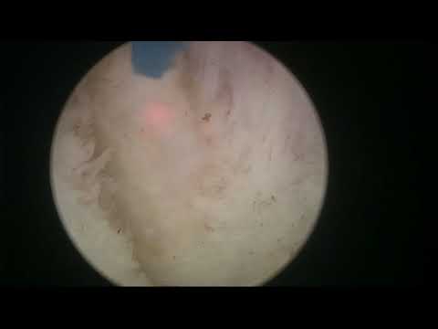
A HoLEP case with a difficult-to-distinguish plane between the adenoma and surgical capsule. The description focuses on judging dissection depth and navigating around the adenoma when that interface is unclear. No particular size or postoperative result is supplied.
Documented details and sources
Only details explicitly stated in the sources. Missing information does not mean it did not occur.
- Difficult dissection plane
Real-life HoLEP 20: Difficult dissection plane
Source checked: 2026-10-06.
Available transcript · English
Automatic YouTube captions, not clinically reviewed. Names, technical terms and numbers may contain errors.
They may contain transcription or translation errors; check the explanation in the video.
- okay so here we are this is a man with a medium-sized prostate you see he has a very high bladder neck and maybe some middle of growth and we're going to do a hollow this bladder is certainly trabeculated and here we are so i'm going to come out towards the apex it's interesting there some stones and some crypts there this is the sphincter edge let's fire here at 12 o'clock i want to mark the white line the limit of the sphincter with the apex i'm going to try to deepen this what i call it the white line a little bit so we can let's say carve a [Music] groove between the sphincter and the element we are so let's deepen the incision a little bit especially uh there's a stone that's why we see those
- reflections of the light in the stone let's fragment it until it's gone so i like to cut this fibers in the posterior area you see here maybe towards towards nine o'clock in this side there's still some piece of the stone okay because this is a way to detach the dissect or detach the sphincter from the apical lower part of the abnormal huh so here i like to cut these fibers see the direction of to the barrel and above because we will try to connect here and dissect i find that find the nucleation plane we can do this gently pushing the abnormal you see that's a that's the nucleation plane and the fact that we did this we cut this fibers prevent damaging the sphincter in this level you see we come
- out you can see the center is completely safe and this side let's enter the plane as well we can do energy dissection we can do a little bit of mechanical uh pressure on the normal to see if the anatomy shows itself you see and we find the plane okay that looks like a good plane to me this is above the rivero there's not much tissue here although we found probably an entry column let's go over the utriculum try to connect the lines the line of attack on both sides i'm going to put my fiber at 12 o'clock and see if i can touch this a little bit so we can start progressing the posterior dissection here trying to follow the white line rhythmically from side to side gently trying to look for the proper
- plane the energy should be able to open the plane without the need of contacting with the tissue [Music] but this plane looks a little bit sticky to me huh so maybe you see sometimes we have to make a little bit near contact with the tissue to catch the fibers that are joining the the anoma to the capsule it's a very fibrous plane very sticky plane all right so we have done this posterior dissection and you can see that the posterior aspect of the normal is totally free from the sphincter but now we have to continue our dissection so i'm going to cut all the adnoma towards 12 o'clock you see i'm going to cut on the enorma this is not the proper plane but when you do these cuts like this what
- you achieve is to detach the apex from the sphincter so this is not the plane we're ultimately going to follow the plane we're going to follow is the the true plane the true dissection plane it's over here but by cutting this initial area where the apex attaches to the sphincter we gain access we gain access and thanks to this access weekend so [Music] so so here [Music] so let's see if i can see a little bit better i have been silent for a while but i don't know why i forgot to to speak so here we have already liberated the apex and now we're going to follow the circumferential line of attack you see i'm trying to position my camera so the line of attack is in the middle it's in the middle of the screen and we
- always have one side of the screen which shows capsule and one side of the screen which shows at noma and you see that we have excellent visibility the plane is a little bit sticky you see it touches a little bit more difficult than usual but this is the thing we never see planes that are exactly the same and that behave exactly the same so we have to adapt with our knowledge and our skills to the individual situation of the patients and try to [Music] complete our work there we are [Music] here we are there we are just following the contour of the prostate we know prostate is a pseudo sphere we have this line of attack to follow you see sometimes the the quality of the flame gives us some tips
- tell us if we are right or not sometimes it's more cryptical you know more difficult to tell but it's very useful to have this line of attack you see because we can follow the line and even in the areas where we're not sure if we are in the right place or not we can follow the line and follow the contour of the prostate which is a little bit you know curved and progress if we leave a lot of unnoticed tissue attached to the capsule we will see that the tissue in the capsule will be more yellow we go through the anoma we might notice uh that the tissue is not so uniform you see the capsule is a little bit of the interface between the anoma and the capsule is quite smooth huh you see so that gives us
- the indication that you're probably in the right plane huh if you see this kind of yellowish tissue here we are more in doubt you see that's probably a little bit about noma or in that enormous nodule you see capsule is more white the norma is more yellow i think that's nice nice concept huh to help you navigate this let's say not so easy planes sometimes but also you see the circumferential nature of the abnormal helps a lot because we are circumnavigating we are navigating around it side to side continually posteriorly trying to adapt to the serious spherical shape being careful here i saw a little bit flimsy tissue here so i went a little bit more inside you see well sometimes you're not 100
- sure that you are in the perfect perfect plane but quite sure that we are reasonably good plane you know sometimes perfect is enemy of the good so it's one of my favorite phrases so sometimes you cannot do perfect with hollow you cannot let's say aspire to have every case beautiful case every case an excellent capsule at normal differentiation you know sometimes you have to accept that things are not so easy as in the teaching videos and books and drawings they're a little bit more real huh real life so here you see maybe it's one millimeter more or less but we are probably doing a relatively good job here it's a little bit yellow so you have to test the tissue a little bit see it looks like a normal see if it looks like
- capsule but overall you know the direction that the capsule and the plane is taking so there we are [Music] going around there norma carefully adapting to the contour here of course it seems like we are reaching the bladder neck area so the plane is going to be ascending and sometimes it's a very abrupt change of direction so i'm going to keep my fiber let's say more close more close to the abnorma not so close to the capsule try to avoid perforating it you see so you can come this way and correct it's a little bit yellow so maybe it's better to try to see if this is at normal or not try to see if we have a good plane or not the plane in this man is not good anyway so but if we don't leave a lot of yellow tissue
- attached to the capsule is probably better here we are trying to [Music] advance in the operation carefully so millimeter by millimeter liberating the anoma from the capsule still yellow well let's see that looks like capsular a little bit deep maybe not critically deep but a little bit into the peripheral zone the anterior fibromuscular zone let's see here we're coming downwards downwards downwards trying to connect trying to see if we're doing the right thing i think this is a good plan yeah sometimes it's difficult so you have to do your your best you can do uh intraoperative ultrasound if you're let's say not convinced or you think that you might be leaving a lot of tissues behind this is a little bit yellow but it
- doesn't look like a very critical amount of tissue there let's let's check check and see if we can remove some more without going deep into the to the capsule [Music] see it's it's it's not very clear but it looks a little bit more whitish here a little bit more white so maybe it's a good idea to continue always very careful of course and there we are this looks better it looks more more white until also here i think it looks a little bit better it connects with the plane over here let's see let's see let's see that's a good connection there here you see i'm firing very close to the anomalous edge [Music] a little bit away from the capsule if i can because i want to detach the normal but i don't want to penetrate
- too deep in the in the prostate capsule up up up there slowly we will gain more access better access and also more insight on the anatomy how it goes how it works if we are going too deep here it looks a little bit deep there let's see it's the vessel a vessel okay let's see if we can enter the the bladder neck i think it would be a good idea to have the reference of of the bladder neck now we're coming anteriorly here this look like circular fibers here you see of the bladder neck so we are aiming a little bit downwards to see we start seeing the change the direction of the fibers see how we can access the bladder this looks a little bit transparent now we might be here now you see these are
- mucosal fibers let's enter the ladder neck and follow the circumference of the bladder neck i think we are quite good here let's check the uo want to see where it is in relation to the bladder neck it's far away see see this side how it goes here it looks like a little bit of a normal i don't know if that's probably capsule here that would look a little yellow to me so it's a matter of finding a good balance between how much tissue you remove and how deep you go into or near the capsule momentum so here this posterior this is connecting now i'm trying to stay here a little close to the normal trying not to get too deep but at the same time trying to remove as much tissue as i can that's posted here it's coming out
- nearly there really close to the to the bladder neck let's see let's see let's see rough posterior aspect connecting already with the sides the other side this is midline below the below the middle lobe huh here we have to see we go so the plane is not giving us any any good information so we have to guess huh yes we know the circumferential nature of the norma we have some reference where we're coming where we're going this is the bladder neck let's see if i can touch all this lateral aspect towards the what's the bladder neck see here if we're coming below the noma see this is looking a little bit flimsy there so we don't want to go there we want to stay up here crossing over to the to the other side
- at the same time trying to connect with what we already had on the other side [Music] yeah let's see no it's trial and error or let's say slowly progressing making things better as we go trying to improve the situation this is ladder neck at the other side [Music] laser is making a strange noise we have heard it before i don't know why it does it okay so where is the uo there huh we are we have a you are a little bit far away from us this is the posterior aspect this is what looked like abnormal you see we went a little bit deeper there let's see if we can release this lower aspect here [Music] elevate it and then try to connect one side with the other side and progressively liberate this from the
- [Music] the capsule posteriorly huh there we are it's not clear huh yeah the plane is not not easy but despite that we are managing to do a very nice uh dissection i think even we took this nodule this is uh here it's the bladder neck let's see this is the other side so let's mark rhythmical line from side to side there we are i'm back see what happens i think we are in a position now where we could elevate the noma and tried to tilt it into the ladder you see it went inside this is a fossa this is the center beautifully preserved and that's almost out but still not very dangerous or critical and this is the remaining aspect here that we have to cut our neck and off it goes i think [Music] this one little fibrous band
- here see if we can remove it let the lights do its work [Music] yo perfect you oh over there perfect and this is the fossa excellent work i think here it's a little bit irregular here this should go i think see what's the fiber breaking up you know it breaks all the time during the operation i want to take this piece out so [Music] yeah the water came out from the endoscope the water uh so this ability was degrading so i thought something's wrong and i looked at the instrument and i saw the water tubing came out of the connector that's it it's a little bit fibrous prostate we don't want to leave any nodule or pad norma behind if we can help it the light has to cut this so be patient until we manage to detach it completely
- there we go that looks much better ah this is uh probably nothing we can also vaporize it with the laser you know these laser close contact and vaporize small amounts of tissue so you have a little bit like this like this like this you can just vaporize it okay so let's most later i think we are have good hemostasis just have to be careful that the catheter will not go in there so typically what i do when i put my catheter inside is to put the finger in the rectum push this part of the prostate up so when the catheter goes in it goes in the bladder okay go this is a plane a prostate without a plane changing the instruments now to to introduce the most later i broke the scope for a moment let's see if i can fix it
- yeah there we are perfect [Music] one of the tops that's it place ladder neck so this is the working position i'm just going to suck with the pedal to engage the anoma and then i'm going to lift lift the tip a little bit away from the flatter neck on the bladder wall and in this position i know that i'm very safe it's not the center of the bladder it's the center of the bladder neck and maybe one or two centimeters inside [Music] the bladder uh except three oh no it got obstructed maybe the tissue is quite hard and fibrous it's not suck it's not sucking [Music] i think the system got obstructed because the tissue is quite hard see it's very fibrous tissue it got blocked maybe in the motor maybe
- [Music] okay so my nurse is very good at uh trying to find out yes peter what's going on yes now there is aspiration there was some tissue inside the blades so maybe that was the the course of the obstruction sometimes it happens as always it's amazing to have a nurse that can help you so fast and who knows what to do because you are scrapped you know the patient you cannot leave the instruments you cannot leave your post you know to to go and check them out later so this is definitely a teamwork operation we are see it's very hard tissue there's nothing to suspect any any problem here just uh sometimes the prostate has some it's a leathery consistence [Music] and harder harder to chew by by the
- morse later see there we are that's the final piece i think we finished this is a nice fossa nice winter let's go