
Surgery
RL HoLEP Videos: 190 cc prostate, Re-do operation with a huge subtrigonal nodule
Dr. Fernando Gómez Sancha · ICUA
Dr. Fernando Gómez Sancha · YouTube
Language: English. YouTube audio metadata.
Surgical content for professionals. Contains footage of an operation.
Playback connects to YouTube. Your search query is not sent to the player.
About this video
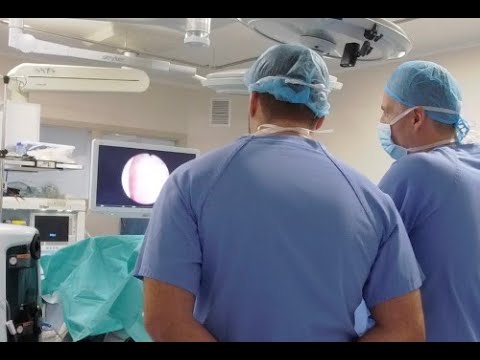
A reoperation in a 190 cc prostate, recorded at Hill Clinic in Sofia. In addition to the previous surgery and gland size, the description identifies a large subtrigonal nodule that was difficult to manage. The earlier operation is not specified in the written account.
Documented details and sources
Only details explicitly stated in the sources. Missing information does not mean it did not occur.
- Redo surgery
RL HoLEP Videos: 190 cc prostate, Re-do operation with a huge subtrigonal nodule
- Prostatic nodules
RL HoLEP Videos: 190 cc prostate, Re-do operation with a huge subtrigonal nodule
- 190 cc
RL HoLEP Videos: 190 cc prostate, Re-do operation with a huge subtrigonal nodule
Source checked: 2026-10-06.
Available transcript · English
Automatic YouTube captions, not clinically reviewed. Names, technical terms and numbers may contain errors.
Selected spellings of enucleation, morcellation and holmium terms have been edited. This is not a full audio or clinical review; the original captions remain the source.
They may contain transcription or translation errors; check the explanation in the video.
- and focus because normally the focus after after more is different than the focus after. So this is the veru. There's a little vessel bleeding there. So this man had an operation before. You see he had a TRP or something before and you see he had a previous operation. So he has you see some tissue was rected.
- So >> yes, >> he was he doesn't remember. >> He doesn't remember, but I think this is uh not a normal not a normal anatomy. I think he had something done before. Let me check if I can stop this little bleeding here. Maybe here also you get an opportunity to see. You see this is the sphincter. It's very difficult. This is the veru.
- Here's the sphincter edge, I think. But here comes all the way down here. I think you see this is all remaining adenomatus tissue. Let's see if I can focus a little bit better. So it's going to be an interesting case.
- This is my white line. This is the recognition of the sphincter in this side. And here is the other side. So we're going up this way coming. Uhhuh. And that's uh you see the veru there.
- Yeah. I think this this is the limit in this side. Let's see. a little bit asymmetric but because here could be well let's see and then the proper limit might be below here like yeah coming down this way you know rather um something like that and it's more more symmetrical more more interesting. Okay.
- So let's see now I'm going to go close to the ver montanum trying to get to see the the plane lateral to the ver montanum enter the paracolicular space let's see here here I will try to do the same apparently Maybe here. You see that's a good one. This is a good one, too. Here we probably coming up this way. Okay.
- Here we have a nice opening of the plane and here we can cross. This could be the ver montanum and so we cross over and connect to do the uh posterior line. Huh. This is a very interesting case posterior line. So we go from side to side trying to develop the posterior plane.
- It's a very nice posterior plane. It detaches very nicely, very easily. I'm not even, you know, doing anything. Just uh Okay, so that's posterior plane. Let's see how further further can we take it.
- It's yellow. So it's probably nodule here. Okay. So, we did a lot of the posterior liberation. Now, let's see if we can um release the apex here. There's this nodule here that probably needs to be removed also.
- So maybe I'm correcting the line of dissection a little bit. The line of the the white line. No, here to come over here. Okay. Let's see if we can enter the lateral plane here. Slowly, carefully, initially lower the lower part of the lateral line to connect with the posterior.
- Here we want to connect this with the plane that we had here. Uh there we are. You see it looks quite good there. And uh here as we come more close to the white line, we need to see where does it want to go. Nowhere.
- Because the anatomy is quite distorted here. It's not so not so easy. Let's try to protect this sphincter part by cutting the aical attachments first and then continue the development of the line. Here we are.
- This is one of these cases where you need to be careful, creative, confident because it's a very strange case. Go. Mhm. Here we're coming up following this this way. So let's mobilize even more coming further up to get progressively more and more mobility and detachment and understanding of the anatomy and the plane and everything.
- Here we are. This is the beauty of Enblock. You know, if you have to do these cases with an incisional technique, I guess it's going to be much more difficult because you know, you cannot even enter very well towards the bladder neck.
- Everything is irregular. So, you know, anything is possible. But I think and block is good because when you finish the epical liberation, the rest is going to be the same as always. This is going to be going around the plane. Going around the plane and trying to touch everything. Let's see what do we achieve here. This is lateral line. I think lateral line.
- Here there is some adenoma. No, here we're leaving it there. So, we have to go to search for it and remove it. No, down here. You see, I took a nodular nodular plane rather than coming all the way capsular. So let's see how can we negotiate this.
- Yeah. And I have to go further up because that's that's a no do. Huh? That's BPH tissue. Okay, let me see. It's now coming towards 12:00 here and the connection to the other side here.
- Okay. to go around this and around uh all that I think try to stay capsular. So now there's a little bit of cows here. There's not a single line, you know. So I'm going to try to do my best to carry this line and try to make a new line that is going to guide us, you know, so we don't confuse ourselves with many different lines. Let's see here. We're coming out very close to the to the capsule. So this is a good this is a good plane up there.
- Let's see where this is taking us. Up here. Yes, it looks good. More or less good. Up there and around. So in order to to do this more complicated cases, I think you need to have a lot of skill and a lot of confidence. And if you think about it, we have salin for irrigation. There's no bleeding. So the patient is it's perfectly fine. It's a matter of finding your way, you know, in such a different anatomy. You know, it's like operating a an extraterrestrial. You know, it's not the anatomy we are used to. But in at the same time, we know that the capsular ed shape is going to be similar to the orange.
- We're going to be dealing with familiar, let's say, familiar planes and familiar shapes. So let's see that looks like a denoma here. Here I think I am connecting the posterior line that we had here. You see this is posterior line. It's connecting very well to to what I'm doing now. So it it looks it looks very good to me or much better now. We we can tell that this is a continuation of of the line that we had uh from below here.
- This is a continuation. And now we can follow this line to go around coming down from below here. You see if you see some deepening maybe we need to get closer to the side of the adenoma so that we don't go excessively deep but this is a plane that we can we can let's say detach very nicely and then here let's see what we have now when we come anteriorly we have all this anterior detachment done and here we are let's say connecting to to the other side here.
- Here's the other side. We started down here. I wanted to protect this uh let's say line of uh mucosa. Let's see if we can because we will need to remove all the tissue that we find here down here. At least this lateral line on the other side coming up and down.
- coming up and down. That's now connecting very well to the posterior line here. This is probably the sphincter edge here. So, let's It was very difficult to recognize. No, but now I'm starting to understand how this is working and uh I feel a little bit more confident that this is correct because we are removing all of this is adenoma hopefully. And uh I'm working on these lines you see connecting the anterior the posterior trying to get a uniform dissection line and trying to connect incredibly it's not bleeding h it's a very very nice tissue probably not bleeding too much here. This is posterior now. Posterior line.
- Now we're coming up here. Now we need to see anteriorly here. What do we find? That was beautiful plane. Now here is the apex. This is the anterior. That's the plane from the other side.
- And this is the connection I think. So we could go up here. Trying to connect, you see, anteriorly with the other side. So anterior line connecting to the anterior line of the other side. So now the aex is uh the sphincter is free from from the aex and we have to deliver due to I mean if you look at the length of the lines you can tell it's a very very large gland.
- It's a very large gland. It was estimated to be 190. So you know probably very very large. Here we have a discrepancy. Let's see we can fix it. So we come. This looks very capsular. So let's just remove this nodular tissue from this side here and connect to the previous plane here.
- So we have a nice lateral line and we can remove all that uh let's say BPH tissue. Many times when we do a retreatment you know it means that the patient had a previous treatment and uh it failed.
- know that because we have the patient again on the surgical table. So when we do a retreatment, we want to try to do the best possible work so that he doesn't need to come again. No, how many times do you think it's acceptable to to treat a patient?
- This is again another uh of the premises of the industry when when they defend the ejaculation preserving procedures. No, you do an ejaculation preserving procedure and you have a higher risk of having to retreat the patient. No. And then you see people presenting cases, you know, I did a 19, it failed. I did a year lift, it failed. So I did a second year lift and then finally he got the final treatment. No. So how how much of that is is makes sense. No I don't know for me personally it doesn't make a lot of sense and I have to say that when I see patients that need reoperations they are a little bit frustrated. No, they they would have I don't know. There's a lot of regret sometimes in this choices. You know,
- patients choose something that sounds attractive. You're going to pee very well. You're going to keep your ejaculation and everything will be like before, just you will be peeing much better. But then they realize that the symptoms do not go away completely. they still have you know bladder hyperactivity symptoms and they end up seeking treatment again you know and then there is some regret and some so yeah if if you can do a nucleation I think you you have to of course we have to listen to the patients we have to see what they want but also we are advisers for the patients and sometimes they get carried away by the pressure of industry you know these companies uh that produce minimally invasive treatments
- for example Tellex revenue yearly revenue is $3 billion you know so it means these companies have a lot of money a lot of money and they can use all sorts of uh marketing tools like you know posing in the forums as patients, happy patients, you know, trying to sell.
- I don't know. I don't know. I think there's a lot of u urologists who start doing these treatments and then finally they leave it for the very, you know, very selective indications because it's not so good as they want us to believe. No. And so universal as they want us to believe.
- For example, I was talking to one guy from Boston and he was telling me ah resume is so good but patients complained. So we have done an application. So the patient has this application and the application explains the to the patient what to expect each day for a month. No.
- So that he can be more relaxed and he doesn't call the doctor all the time, you know. So it means that when when the doctor gets called by a patient all the time, you know, I have pain, I have discomfort. When is this going to get better? No, you told me it was minimally invasive, but I'm not feeling that it's minimally invasive because I have a catheter.
- you know, so if if they have to do an application to explain to the patient what's what's going on it means that uh it's not so nice and of course when doctors get a lot of calls they get discouraged you know they think well this is not worth my while no because here we are this bladder neck which is also modified by the previous operation.
- I think it's a nice nice last case for the day. Yeah, very beautiful. Very beautiful. Very interesting case. >> But as I said, if you if you of course you need to understand the anatomy, you know, the more cases you do, the more equipped you are to to do this stranger difficult cases. But you see that uh we are doing the same we are using the same principles of the operation. We are using the similar you know strategies and uh it's just tailoring it to to this particular case which looks a little bit more difficult, more demanding.
- I think the aics dissection was a little bit more tricky. The image of the sphincter was not so clear. But then of course the sphincter begins where the ends you know at the end if you if you see the tissue of the adenoma if you see how it gets to contact with that area then you you understand better. No sometimes in the normal anatomy we can distinguish the sphincter very well. In this kind of anatomy, you know, sometimes we have to do a more progressive careful initial approach and then slowly get to understand where's the where's the sphincter, where's everything and try to navigate this these planes. No, on this here we're coming up.
- This is looks like capsular This looks like good a good plane. Let's see. That looks yellow there. So maybe a little bit of aoma remaining there. trying to go around millimeter by millimeter making sure that you know we don't see perforations.
- So if if we see deepening in the capsule we have to correct a little bit the depth of the dissection. But if we can take all this tissue out it will be much much better I think for the patient. Now we have to remove all that bulk of tissue.
- Now this is the connection of the posterior plane with the lateral plane here coming up aiming towards the bladder neck more anteriorly. This is a nice case, huh? Very complicated anatomy. Crazy. [laughter] Crazy. Yeah. Yeah. Well, at the end it is an extreme case, but you've seen now what 15 20 cases and almost 20 cases and uh now you can see what I'm doing. You can understand what I'm doing.
- And you will and then my recommendation when you do a a retreatment case sometimes you see that one lobe was very well resected for example and the other lobe protrudes over the you know and looks obstructive. So many people think if I remove the remaining lobe it's going to be okay. But my recommendation is to always go to the plane in the Veru, you know, do uh and block exploration of the plane because many times you get people are very confused and very impressed because they see that even in the area that looked very well resected, you know, there is a lot of tissue remaining. So when you look for the plane, when you go under this properly, you know, apparently properly resected place, there is a big big big chunk of
- tissue there that could you can remove. No. So that's because the endoscopic image is not not always um not always uh telling us the truth. You know what I mean? >> It's it could be somewhat misleading sometimes.
- So here we're trying to come down. It's it's a very very big prostate. And I think this man is lucky that we were here today to operate him because this would be a challenging case for any endoscopies. Don't you think? I mean, if you do a resection, maybe you can get a bigger bigger hole.
- What's this? Why I'm I experiencing some difficulty moving because there's too much bulk. So, this would be posterior plane. Posterior plane now getting closer and closer to the region of the bladder neck.
- This is the lateral plane. We now we we have to start getting closer to the adenoma because we want to you know the the the plane of the capsule is going to become more perpendicular to the fiber. So we are more careful now to get the energy closer to the side of the arenoma than to the capsule you know to prevent deepening in the capsule and perforating.
- Although this man has a very very thin capsule. Very thin capsule. It's his bladder neck. I think bladder neck. So here we can go like this. >> What about this? >> Yeah, we'll have to check. Let me let me go there. I want to reach this four 4:00 more or less here. You see initially I cut the bladder neck but when I get let's say to 4:00 I I will check you were right because we are probably very close to to the UO. Here it is. You see but now it's safe because the edge of the dissection is here. So we are okay there. You see this is now the fossa.
- It looks very nice. Anteriorly we opened as well. Now we need to focus on this side. See how can we so you see we're going to give this man a simple solution to his uh how do you say complex problem. No, because this was a very large I mean you could do an open procedtomy or something but we have been able to you know find the the right plane and we are going to deliver the adenoma completely. So hopefully he he doesn't need any more treatment in his lifetime.
- It's posterior plane down here. Maybe we need to find it. Find the the right plane to to follow. We see a lot of yellow elements. We want to [clears throat] Okay, here we are. I think it's a reoperation case. The Dr. Schumanov said he had no previous operation, but I think he might not even remember, but it looked as a redo case clearly to me.
- Here we are coming connecting coming up and around the the trying to follow the nice plane posteriorly. Remember to stay aim aim towards the adnoma. Huh? Because it might have a very big uh retrotrional component as well. You see that looks like a big big nodule.
- Here you see this looks like a big big nodule here round. You see that's probably going retrotional. Let's see how can we negotiate this dissection carefully. Let's bring this plane that we had here.
- Come connect here to this side here. Let's see. Now this is the bladder neck in this side. So it's wise to cut the bladder neck in this side before we go posteriorly. No, because we will have much better orientation like always. Like always coming up towards the bladder neck.
- Now that's looking slightly deep. So I'm going to stay a little bit closer to the edge of the adenoma here. So here we need to see the UO here. It's lateral to to my line. So we are okay.
- Let's see how can we connect this below to that above. Let's see we can you know negotiate this curve here. That's a seminal vesicle. We need to keep more superficial. What is that? That could be the retro trional space. Let's see here also. It looks very deep. Let's stay up.
- That's also looking deep. You see, but there's a big big nodule here that goes probably below the below the the trional area. You see that it inserts itself there. Stay always close to the the nomato side.
- Minimize if I mean small perforations as I say always are allowed. You you can do a little perforation but you shouldn't be doing the big ones. No. Here again we need to stay inside of of the plane. So we go cutting more even more closely closely to the side of the adenoma to see if we can remain inside the the capsule here. It jumped a little bit. So that plane would be crazy to follow. But here you see we managed to correct a little bit. So now we can continue working.
- It jumped again. There is some tissue here that when we reach there, it jumps. Let's see if I can stay close to the the side of the adenoma here. This is from up to down. And this is from down to up here.
- Here. This was the good plane. And it jumped again. Probably we are trying to develop this a little bit more. You see, we have to do small improvements. a small small improvements because this is the bladder neck and the uo we saw it was lateral to to this is blad neck as it was resected before I think so here we can also mark from above we could go and see the other side see how it looks like here bladneck the yo looks a little bit funny. No, this this tissue the UO was uh it looks like a papillary condition. This is the UO and here from the anterior you see we can see that this is the line we did. So we could bring it here to connect. Huh? So we can go from above as well and uh connect a little bit. So when we reach
- this point from below, we are already there and there is no danger for the for the UOS or anything. Okay. Also, we need to see how mobile this this uh piece is because that is getting under I think when you look at the this bowl, you see it's going under the trion here. We're gaining some mod mobilization from the side and from above.
- Let's see curve. Crazy crazy anatomy with this man. Let's see if we come from this side. Maybe we can mobilize this lobe in order to be able to push it into the bladder. So we do minimal minimal improvements, careful improvements, trying not to go to do something horrible, you know, just to see if we can negotiate the release of this big big nodule here which is going retrotional. I'm think I hope well I know that we're going to see a huge retroonal cavity space lined by a very very thin capsule in some moment.
- Let's see also if I can if I can tilt if if the adenoma will tilt into the bladder. It's not so not so easy. Not so easy because there is still a lot of Look at this beast. The more we liberate, the more we can move it.
- So finally, I hope we can get some. You see that's the capsule. It's like a membrane. What was that? Some water stuff. No. So I stay I target my laser against this here. Maybe I can push this side of the prostate or this lobe, you see, into the bladder. Let's see if it will want to go or not.
- It moved a little. It moved a little. Now when I push a little bit more, it goes in the bladder. And then I hope that we have much more space down here to reach there. You see this ball of tissue is in the retro trional space. So I've seen this beforeh is is some some it grows below the bladder the adenoma it goes very very deep there would be nice to be able to negotiate ah the passage of this lobe but it's too big and it doesn't move air when I push it So, it doesn't want to move. Maybe not yet, sir. Still too attached, I think.
- Down here. Look at that. It's absolutely crazy. Here we should see bladder neck. You see? And below below the bladder neck we have this ball of tissue that is deepening there. So here we are reaching the bladder neck level.
- Also detaching some of these crazy crazy attachments to the blad neck. region here. Absolutely crazy. Okay. So, slow progress, but somehow we are getting closer and closer to the edge.
- And the moment it's free, I think we will be able to to to tilt it or to move it. Now it's moving more. I think it's lifting. Not so easy. There's not so much uh more left, I think, to release here.
- This is nearly nearly released. But it's such a big protrusion. It's like a nose going inside the you know because we could lift it. I think we could be pushing the everything inside the bladder.
- You're getting there, huh? Now it's it's feeling better. It's moving better, I think, too. Let's see if we finish with these attachments here. That's the nose. And I think everything went.
- Now we need to do some hemoasis and see because the cavity is huge and there must be some little bleeder. Let's do some hemoasis and see what what happened here. You see this is actually absolutely retroonal. There is like a cavity here because this was this this is the footprint you see of this nodule that is under under the trion here. Here's the trion. We're just growing into that like a big nodule. Huh.
- So you see we can achieve a lot by using the energy carefully and uh I don't think it's perforated there. I think it's just a very thin stretched capsule. You see? So it was a very very very interesting case. We went almost out uh in several occasions, but you can control your action and uh try to correct the direction of the So keep keep the little perforations small.
- So crazy crazy case, huh? Very very beautiful. But as I said the same degree of uh happiness I mean when you have a rectocope in your hand you feel very very well equipped you know and uh you feel that you know what you're doing you know what you can do you know uh I after after you get experience with the laser you can also get that kind of uh feeling no here when I was pushing probably I opened the plane a little bit sometimes the The plane is so so thin that we can break it uh mechanically rather than with the laser. I didn't do this with the laser. Probably it was more a mechanical uh tear. Huh. It doesn't look bad. Probably having had an operation before. I think everything is fibrous around the prostate.
- You can see a little bit of fat here. This is a, you know, moderate concern in the sense that we're trying to morelate now. So, it won't take long to to finish hopefully. So, this is the huge huge huge fossa that we found in this case. And as we go out, you see, we're going to see that uh probably this is the sphincter. You see this is the sphincter. This is the sphincter's mucosa covering the sphincter. The ver the ver is quite forward there quite far away because we um of course it's it's it was a very very large prostate. So I'm going to leave this here. Try to stop the bleeders because we want of course the patient to be safe in the post-operative period.
- Here we left a little bit of uh you see we mistake we did a little mistake with the plane. Uh we believed everything was removed but there's still some. So that's why this phase of hemoasis and trimming is useful.
- We can correct the little mistakes we do. And if we leave some tissue, we can take it out while we also do hemoasis. He stasis. And I want to release this little piece into the bladder.
- It's a crazy case. Okay. So, there we are. I think the outcome is going to be excellent. Patient will pee like a horse and I hope that this will all contract in the post-operative period and we can remove the catheter soon.
- Hopefully he will be continent. He has a second bladder here. >> Yeah. Maybe use a catheter for apply the For longer, for longer. Um, >> no. >> No. What you >> insert? >> Say again.
- >> Will you use the catheter for the apply the fo >> attraction? You mean no f? >> Yeah. >> Oh, you can use a flet. >> Put the put the flet. >> Yeah. Yeah. We put a fo catheter. Let's put a flet catheter. I will use my finger to to lift all this. So the catheter goes in the bladder. Huh.
- So maybe the introduction of the catheter would be difficult in this case. So let's see we will be careful so that we don't leave >> the catheter prefer the >> maybe maybe >> it so I'm asking >> it's possible it's possible you know we we usually get very good hemostasis so hopefully he will not bleed and then where the ca the catheter is positioned is is irrelevant it doesn't matter where it is okay that was this is all the a little bit deeper area here.
- Look at how how it went all this way under. Mhm. Okay. Let's try to morsel it. She's okay. More later. Let's focus on the tissue here. Let's position the blades over the bladder neck here and let's start midline of the patient and hopefully we can concentrate on morcellating all the time.
- We don't want to stop. We don't want to the water is open. It's entering the contact of the tissue with the Morcelator blade is good. So, let's see how long it takes to remove the the tissue. But I hope we can be as fast as possible.
- Let's see. So, it's 400 p.m. now. We have done eight uh holyps today. This is the last case of the day and it's 4 p.m. We started at 8:30. So, we we're going to do eight cases in 8 hours.
- So that's quite quite good because it includes the changing time from patient to patient. So it's really really an efficient uh work here. Nephoscope is a little bit uh degraded. I think with the use I think it's not so it doesn't let us see so well anymore.
- we can see enough, you know, but sometimes there's a difference between the the view that we get with the one lens and the view that we get with another lens. Also, you know, morcellation in when we the settings of the morcellator are also important.
- There are two factors that we can modify in the console of the morcellator of the pirania. One is the rotation speed. Rotation speed means how many times it rotates um you know in the inner blade, you know, around the or over the the the outer blade. No.
- And I'm I'm using 1,500 revolutions per minute. Okay. So, it rotates pretty fast. If if you make the math math, if you divide 1,500 times 60 minutes, you know, you will get sorry seconds in a minute, you will get how many rotations you have in uh in one second. Okay, so in one second I think it's something like 25 or something like that because if you multiply 20 by 60.
- That's 1,200. No, it's like 25 I think 24 something like that per second. So the blade rotates very fast. Um there's another factor that we need to set and this is the frequency. The frequency as you know is the cycles per second.
- So basically we have to tell the morcellator how many times per second we want the blade to run a cycle of left and right movement. Okay, one cycle would be to rotate to the right and to rotate to the left. So for example, if we use one hertz, this is going to be 0.5 seconds to the right and 0.5 seconds to the left. Okay?
- If we use two hertz, which is what I'm using, it's going to make two cycles in one second. So, right, left, right, left in one second. And this is the rhythm, you see. So, maybe we're dividing 25 in uh by uh four, you know. So it's uh about six rotations to one side, six rotations to the other side, you know.
- Uh so in 1 second is about 24 rotations per second. So it rotates really really fast as as we saw yesterday. If we have very hard tissue, we can reduce the speed of rotation to moreellate harder tissue.
- But so far so good. I think the moration is quite good. I enjoy very much these cases. I was telling you today, you know, I hope we have a retreatment case because um they are very beautiful.
- And as I said always, always always irrespective of what you see in the endoscopy. You think you know the left side is very well resected. Go around the ver look for the plane and see what you can find there. You know most of the times there's a lot of tissue and if you clean the the fossa completely you know this tissue will never grow back and uh you will save uh the patient the possibility of an third operation because you know now people live a lot live many years and typically you know with TRP we operated all the BP PH studies on surgery the mean age is 60 65 something like that so you know a 60 year old man today could live to be 85 25 years you know that at 10 years I think after TRP there's like one in4 uh chance of
- having a reoperation so you're going to have a lot of patients that when they're 80 when they're 70 75 they will need a reoperation for their BPA symptoms So if you can treat them when they're 65 and remove everything, you know, I'm seeing patients I operated many many years ago, 20 years ago come to see me and say, "Look, I'm doing well. My prostate is 15 grams. I still pee well, you know." And to that man, I mean, he went to the operating room once and uh he peed well for his life.
- And also I tell the patients that uh the prostate complicates their old age sometimes. So many many people old men die because of infections, catheterss, stuff, you know, because their choice when they were 65 was to do a partial resection of the adenoma.
- So I'm not blaming anybody. I'm just saying I feel well doing this, you know. Okay, change, change. You know what I mean? I feel well when I do this kind of work because it's giving the patient a better chance. No, you probably got like 80 g out, something like that.
- It didn't come with a catheter. No, no catheter. >> No. >> Well, the center is is >> ah maybe the catheter. >> Yeah. When when patients have a catheter, it's like wearing a splinter.
- You break your arm, you know, and then the muscle gets atrophied. So I think when you have a catheter for a long time the sphincter uh probably experiences some atrophy because it's never closed you know it's always open because of the catheter so but I have operated here I have operated patients who had a catheter for many years 5 years 6 years 10 years extreme cases no and uh the patients were continent right away or or very soon after after the enucleation socred Incredibly the sphincter works seem to work despite but if I have such patient I tell them you know maybe maybe you will have some stress incontinence for some time if you have a hypotonic sphincter and maybe some rehabilitation could be a
- good idea I don't know we see all all kinds of things that all kinds of surprising things sometimes And it's difficult to generalize. You know, some people enjoy let's say categorizing different subgroups of patients and sometimes they take it to the absurd. No, in these patients with 80 gram prostates who take finasteride, I have to say that in my experience, they behave like this or this happens, you know, and you think why why do you do that? But I mean it's it's very difficult to once I was asked by a colleague have you noticed that patients with catheterss have better planes.
- I don't know what the catheter does to make sure that the plane is better and I told him have you considered that the patients with catheter probably have bigger prostates probably. You know what I mean? So when you when you take this this personal experience and you try to make conclusions based on your casuistic you know your cases uh it's very unlikely that you're going to be right.
- Uh so I prefer to say you know every prostate is like an orange. We're going to peel the orange. Some prostates will peel very well. Some others will be more difficult but at the end if you peel the orange uh patient will be happy. No, most of the times or at least if you're going to do this kind of study uh trying to establish correlations I mean it shouldn't be based on your personal experience and memory.
- It should be based on a more you know maybe statistical study or something. So nice case to finish the day. Let's see if we can morcellate all this tissue and then put the catheter. The catheter might be a challenge because of that retrotrional cavity is removing this.
- uh >> normally I would try to remove the catheter tomorrow but here they tend to keep the catheter for a couple of days. So depends on when you are. I I am very aggressive removing catheterss because surprisingly cases like this sometimes pee very well.
- They go home and there's nothing wrong with with them. You know they can empty more or less with these patients. You know when they go to the toilet if they relax the sphincter the urine falls out.
- you know it's so it's incredible that you don't need long periods of catheterization and you get more and more uh confident you know sometimes when I did a case like this who had a you know a rough area where there is some fat visible like that I thought I will remove the catheter tomorrow and then or maybe I I wanted to remove the catheter the next day but then when I go to see the patient in the hospital, the nurses tell me, you know, we do took the catheter out. Oh, I was hoping to keep it, you know, but then they do well, you know, and then of course you you start to get more and more confident removing catheterss early, you know, because you see that for the majority of patients, it's it's okay.
- I think with this next day removal of the catheter, we get about 5% retention rate. So this these patients will need need to have a catheter back in maybe wait another day or two and then remove it.
- After two or three days I think nearly 100% the patient's pee. You have to make sure that remove to remove the clot in the fossa before removing the catheter because clot retention is qu one one of the causes of sometimes you know if you don't wash out the fossa properly before removing the catheter some patients can have a retention because of that one minute okay change okay let's change the bucket again.
- Yeah, we'll go to another 50 out maybe. It's a big one, huh? It's a big big prostate. >> I suggest any specific exercise. >> Any specific >> exercise? >> Exercises. Normally, no. Normally, they don't need I I tell them to take it easy for a month. just walk around but not uh do exercise and uh to avoid sexual intercourse because uh they sometimes they want to experience the retrograde ejaculation and they engage in sex in sex very very early you know and uh I tell them that if they go very early >> they might experience pain, irritation, bleeding Okay.
- >> So, but if if you want to uh if if they have some leakage or something stress incontinents something like that, >> they can do kel exercises but not too early. I think if they start doing exercises very early maybe they will irritate all these wound maybe they bleed more.
- So we have also this magnetic chair you know the MCA. >> Mhm. >> And uh we don't use it too much I have to say because we don't get a lot of stress incontinence. So, but this magnetic chair apparently is very efficient more than the self uh practiced uh contractions of the pelvic floor.
- >> Say again. So, it's working well. The tissue is coming to us. I think we are nearly finishing the morcellation. So a very interesting case. There we are. So all the tissue came out. I think I have to check for fragments.
- Try to insert a catheter carefully. Now there's nothing in the fossa. So here sphincter. Thank you Petia. Let's put the catheter with carefully. You feel like a horse. The man has a big hernia also here. So but it didn't bother us very much.
- Mhm. Let me put the finger. Try to get in there deep enough to push it up. Okay. Push all the way. Yeah. Fingerh. >> Chop yourself. >> Say again. >> In Turkish. Chop yourself. >> Ah. Okay.
- Here we are. Let's get uh 60 for example.