
Surgery
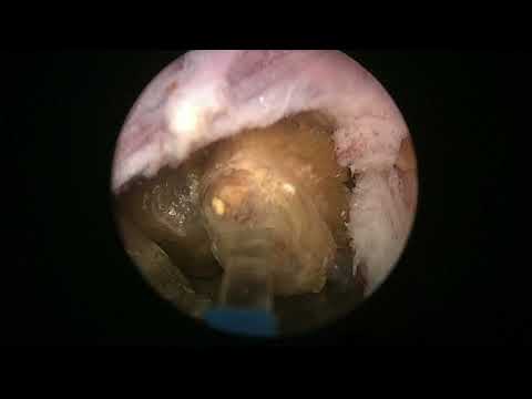
RLHoLEPcase 32: Stony prostate, unusual case, not en bloc this time...
Dr. Fernando Gómez Sancha · ICUA
Dr. Fernando Gómez Sancha · YouTube
Language: English. YouTube audio metadata.
Surgical content for professionals. Contains footage of an operation.
YouTube applies an age restriction: if playback is blocked, open the video on YouTube.
Playback connects to YouTube. Your search query is not sent to the player.
About this video
An unusual HoLEP case described in the title as a stony prostate and explicitly not an en-bloc procedure. The author identifies the video as an unedited, full-length recording with intraoperative findings. The written description does not specify those findings further or explain which alternative dissection sequence was used.
Documented details and sources
Only details explicitly stated in the sources. Missing information does not mean it did not occur.
No clinical case details have been extracted for this video.
Original title and description on YouTubeSource checked: 2026-10-06.
Available transcript · English
Automatic YouTube captions, not clinically reviewed. Names, technical terms and numbers may contain errors. Check them against the video; complete audio coverage has not been verified.
They may contain transcription or translation errors; check the explanation in the video.
- hi this is fernando sancha with another real life holocaust this was a strange case a really particular case i went to the south of spain to do a list of patients in the waiting list for the national healthcare system and i had this patient who was performing self-catheterization because he had very large residual urine volumes and um it was incredible that after performing self-catheterization we found these two consecutive rings in the urethra they don't look i mean the caliber doesn't look very tight so probably not not the cause for the for the
- obstruction uh and and this patient had a urodynamic study and he had an uh previous spinal surgery and after these surgeries when he started developing the problem so he had some mention to a urodynamic study where they said that maybe he had a dysfunctional voiding so it was all a little bit confusing and so yeah probably whoever indicated the operation thought that he might benefit from removing the adnorma he might have a easier self-catheterization or he might [Music] improve his emptying without catheterization so
- this is uh what i found initial uh two rings in the urethra and then a relatively small prostate not too big a little bit tight in the bladder neck and i hit [Music] very very uh bad looking bladder there was pus there that i emptied uh so one of these peculiar cases that you you can find uh it really looked like a neurogenic you know bladder shape going all the way up very high capacity but uh high you know trabeculation so i was going to start the
- classical and blocking nucleation and i was marking the limits and then we found some stones some stones so probably uh due to the you know strictures in the urethra i always think that this strictures in the urethra can cause an increase in pressure in the prostatic urethra so the bladder trying to contract trying to empty the urine refluxing into the prosthetic cannonicles and ducts and [Music]
- dilating these ducts and then of course having this sedimentation and lithius formation stone formation so i well i was trying to do the unblock technique but i i found that being such a small prostate the drainage of the water was not so good um often the sphincter contracts uh over the emptying uh orifices in the in the the shaft of the um endoscope and uh yeah it was very difficult uh to to to to progress there so i decided to do a more classic approach where i would probably do
- incisions you know remove the middle lobe area at the beginning because it was not so easy to to to work with all these stones hitting the the lens and with relatively bad visibility so for a moment i was thinking how how am i going to do this so yeah you see it was almost impossible to to progress so of course when you find a strange situation like that you have to think a little bit remain calm and uh you know just find a solution uh there were many many stones of course when you want to remove the prostate you
- want to remove all these cavities so not only the stones but also the the cavities where they formed so here i think maybe i was thinking you know what to do where's the uo here and i decided to do a more classical approach so trying to carry out an incision in one side of the bladder neck this would allow me to wash out all these stones i hoped so here carrying down the incision to me it was also always a mystery you
- know and i was doing the three lop technique uh that's you uh it was always a mystery to know when to stop with this incision so uh that looked like uh the frenulum of the arrow there so i thought maybe if i fire a little bit i will break these stones into smaller stones you can see some kind of popcorn effect but the truth is when the scope was at the apex it was not easy to see to see much all right so i was going to start the second incision so what i was saying is that when i when i did the three lobe technique i usually try to find uh a nice steps in the
- incision but then i would check near the veru where the proper plane was and then carried this plane upwards okay that's the other you all the the uos are going to be very very close here and this is another incision here i'm cutting on the on these cavities and all these stones yeah i have seen a number of cases like this in my life and some of them with hundreds of stones inside the prostate if you do a rectal examination it feels like a bag of stones and yeah it's quite uh quite interesting so
- you know things are quite relative i've if you go in and you see the strictures like that you might think the structure is a problem so what should i do should i touch the prostate or not maybe maybe this patient should have a urethral reconstruction so sometimes you have to judge clinically what to do and it's not easy to to decide [Music] what is the best course of action of course normally in my practice i i would have done an ultrasound and i'm sure that i would have seen a lot of stones in the in the prostate and then probably that would have prompted me to do a transactional ultrasound or or some other examination you know to to find out but uh this is what i found and
- [Music] so that i tried to communicate both lines more or less in the midline you see working in the apex was quite challenging because of these stones [Music] bothering there you see that's the lining of the cavity and i want to go deeper than that because i want to remove the cavities so i want to remove this these cavities are usually formed inside the prostate so we want to go deeper than that so the incision has to cut through the floor of these cavities as well you know because you want to remove the whole tissue there so this was the progressive approach the nice thing of using a laser is that
- usually the bleeding is quite controlled so everything you do is accompanied with good hemostasis so this way you can you know gradually get where you want to go it doesn't it doesn't uh you shouldn't be anxious when you when you do a strange case like this and there are many strange cases with with the bph when you when you try to go to the plane you find interesting interesting things sometimes i have found abscess cavities gold abscesses you know maybe filled with bus you know going below the bladder with very high capacity uh you can find a lot of things
- and you have to keep calm and try to think and understand of course it's better to go to the operating room with the patient properly studied but still sometimes you can have surprises so here i was trying to look for the deeper plane where i would be able to remove this cavities with their lining mucosal lining of the cavities i was progressively deepening the incision so trying to get to the proper depth to make sure that i remove this completely one of the well this is the finding that you can you can probably encounter when you treat patients who have had a history of
- recurrent acute personalities sometimes the original cause is a metal stricture or a urethral stricture that is not properly you know evaluated and it's not properly found so they have this hyper pressure in the prosthetic urethra when they void and they develop this dilated ducts where the stones form and of course this is a fantastic environment for bacteria to grow antibiotics do not reach there very easily so yes that was the uo so i had to be very careful to mark the incision properly so to protect the uo let's see that everything's funny in this patient everything is
- strange even the anatomy of the bladder neck was quite quite strange so that's the release of the middle lobe tissue trying to follow the plane the problem also in this case was that the plane was not very easy to see i i couldn't find a proper a proper plane it happens uh often with the small prostates and here you can see how i did this incision initially and then i looked for the depth near the veru and then you bring this depth up because i don't know unless you are very experienced or [Music] it's uh sometimes difficult to distinguish where is the limit to your incision you might go
- too deep or maybe not deep enough so i do this progressive approach when i do a three loop technique of course it's not the technique i do every day so i you know i i i use that that trick probably if you do it every day then you're better at judging when to stop the incision and maybe you get it right from the beginning but it doesn't matter if if you do a deep enough incision maybe not not all the way and then you complete the depth as you travel up towards the bladder neck you see like here deepening it more but with the knowledge of how deep the plane is so this is the quanta system
- laser using virtual basket at my settings of 2 joules and 50 hertz there's a lot of discussion about settings and i i want to stress that core settings are important they're going to determine the output of the fiber and [Music] but [Applause] i think they're less important than uh for stones more or less everybody fires the stone in a similar fashion but when it comes to dissecting the plane there's a lot of variation in the speed of movement of the fiber you see if you have seen my videos i tend to move relatively fast so to do a very soft dissection
- uh off the plane careful dissection of the plane so this but there is a fast uh generally speaking you know fast movement of the fiber along the the line of dissection uh but other surgeons are extremely slow so maybe using the same settings the tissue effect is going to be very different so some statements like you know if you use a low power laser you're going to cause less desuria i would say it depends it depends on on your technique it depends on how close to the tissue you fire on how slowly or fast you move your fiber that by the way could be a agricultural doctor sometimes we see them there's no uh [Music]
- problem if if you see them like that i mean often you cut them near the veru and if the plane you're following is a little bit deep uh into the central zone of the prostate uh sometimes you can see the ejaculatory ducts or even the the differential uh uh amples or dielectricians as the similar vesicles uh get inside the prostate you can see the sometimes even the seminal vesicles and it doesn't really matter ah don't panic if that happens it has no clinical significance that i can that i can find so often often with uh holy the planes are not
- easy often you're following for example a prosthetic nodule and you you know dig a little bit deep to take it out and then you find yourself in deeper regions and it's usually no no problem so don't panic just continue working do your hemostasis and here i was looking for the uo where are you uh trying to set ah there it is it was too close so and now i'm going to mark another line you know because my line of uh incision was a little bit lateral initially to the uo so here i had to correct a little bit to safeguard the uos in both sides so
- here i am trying to cut through this very very muscular tissue very tough fibrous tissue and really wanting to get to a point where all these stones can be pushed into the bladder to to be able to continue with the work so i thought this would be an interesting case to show because it it has several several components you know the initial the initial structures the stone collection and uh the fact that it's a small prostate with a difficult plane
- but there it is i think the the middle up flipped and the uo is there okay so just a little bit ah there it goes there it goes that's the the promontory of of this other uo and now all these stones that have to be [Music] tried to to evacuate them some of them came out yeah typically we put these stones they're captured by the by the gone that how do you say that there is a there is a basket at the bottom of the water collection uh system and uh they tend to accumulate there and then we give them to the patients with a little uh
- a little [Music] jar or something with the stones inside and this is an attempt to do the 12 o'clock incision it was a little bit difficult to reach this is a step i never liked off the three lop technique because in order to reach there you have to push the the scope down to tilt it so hard that often i think we cause a sphincter split at 12 o'clock so i it's very different when you do an unblock technique basically what you're doing is you're accessing the prostate from the side here again you see there's a stone and a cavity and i want to remove it all i want to remove that mucosa also so i i try to see and understand
- you know how far could i go here and try to to remove all these cavities to be aggressive at the apex trying to to find the the lowest possible plane here that's a little bit of mucosa i think the sphincter was quite uh okay at the end but yeah you have to go and take out everything you can like that there's more at the lower epics so yeah let's see trying to remove the apical
- optical tissue on the epical cavities because i think if you leave cavities or [Music] even you know this unfractures mucosa there i think you're going to find that or there's going to be a risk for infections later on so you know take your time to try to remove as much as you can from there and do a nice toilet of of the prosthetic cavity probably i mean in this man he he had no history of infections but yeah i i always feel that it's a good thing to to remove all this tissue and all these stones from inside the prostate
- so this was trying to reach the 12 o'clock uh yeah incision to connect you see i i think if you do a three load technique you can benefit from uh doing the white line at the beginning of the procedure and then you do your three long incisions so when you do your 12 o'clock incision you will [Music] carry it out until you see the white line and then you protect the sphincter and there's no curtain you know the because you you avoid breaking them of course you you avoid ripping the center of the mucosa when you're working so i think before i was trying to say that this split at 12 o'clock that you can do
- when when when you tilt the scope a lot to do a 12 o'clock incision is avoided by the unblocked technique because if you when you do the unblock technique you start the dissection from below and progressively you put your scope on top of the apex and then when your scope is on top what you do is you lower the prostate so when you reach the bladder neck there's really not much tension there and i usually do not see the split at 12 o'clock mucosa caused by the traction uh you know i think that's one of the possible reasons for for uh stress temporary stress incontinence after the three lobe technique
- one of them is pushing the scope too hard to to reach the 12 o'clock region which is sometimes very very high in in patients with big lands and so i prefer this this block approach because it allows you to lower the prostate and when you reach this uh bladder neck you're lowering the bladder neck too so when you cut you don't need to exert so much pressure on the on the on the sphincter so here is the lateral dissection of the lobe trying to connect the lower aspect with the upper aspect and [Music] again you can see that there's no beautiful plane and almost of the cutting is down in contact with the tissue because it's so hard you know there's no nice plane that separates
- beautifully and so i'm doing a little bit of contact dissection there because when you do contact uh of course the disruptive effect of of the laser on the tissue is much much more intense and of course that can help you cut through fibrous tissue inflamed tissue chronically implant tissue like probably this man had very very hard prosthetic tissue there you are also you always think in the oh that's a that's an artery there so let's leave it alone you always think when you operate patients with small prostates that maybe
- the the cause for their difficulty boiling is not so much the bph growth but uh possibly the bladder neck yeah sometimes so i don't know my observation is that in patients who have a um a small prostate and obstructive symptoms they tend to have thicker bladder necks than patients with larger prostates so again this is the other side trying to this lobe was looked smaller so i was trying to find my way trying to find where where the the capsule would go you know sometimes you see a plane everything is very clear and sometimes
- you have to to try your best and this is a recurring thing with with olip hollop is not always the ideal operation you see in the videos here for example you see i i see that we can take this there's even stones there so we can take the line a little bit lower to to dissect but uh with experience the laser becomes your tool you know and everything you could do with our sectorscope possibly you can do with with a laser you know there's no it becomes your tool and then you have to adapt to the clinical situations uh and here what we want to do is of course do a good toilet of all this tissue and all these cavities and all these stones if we can
- and and leave the fossa uh free of all that trying to look for the best possible plane that you can find of course you can distinguish when you go uh a little bit deep you can distinguish the depth that you're going deep you know you start seeing that you're going outside and then you can stop and and correct and uh this is what you use to guide yourself [Music] so if you're not very experienced and you find a case like this i mean it's a small prostate you can always resort to to to their sectoscope but i would uh recommend you to keep calm and try you know keep trying
- because often you can find a good resolution for all these stranger cases just keep calm you know do things that are make sense correct yourself if you find that you're not carrying the proper plane another nice plane or a nice nice and off plane and yeah stop thinking that hollop should be a beautiful perfect operation like you see in the video so it's sometimes it's not as perfect not as beautiful sometimes you find this kind of structure in the urethra sometimes so you have to use your judgment of course when i see a urethra that has a structure that is long and hard
- and very tight then i probably would stop and consider doing a urethroplasty or even doing a urethrotomy and let's see if the aerothoratomy improves the symptoms of the patient sometimes patients pee really well so the prostate what had nothing to do with their symptoms but interestingly enough sometimes i've done blood and sorry incisions and i said said to the patient you know i didn't want to operate the prostate if we're not sure that the prostate is the cause and then the patient comes back and says doctor i i am not being well you know i calibrate the urethra and there is a good caliber after the urethra me but the patient's still
- not voiding well so you know one thing is the guidelines and the recommendations and the other thing is how do you you know face the real cases in in your practice so yeah it's always an interesting question we do urethral reconstruction we do urethroplasties i had the luxury of working with with tony monday in london for for one year one whole year [Music] when i was a resident and i learned a lot with him about urethral reconstruction and we do regularly urethroplastis and we enjoy doing them of course but [Music] different clinical scenarios but well
- this is one case where i found impossible to do the unblock technique so i had to switch so it's nice to keep flexible and to keep open to [Music] see the plane is not there so you have to judge where where you're going and correct once and again until you you get the desired outcome no it was uh it was an interesting case you see the tissue is very hard to cut i i haven't seen the histology for this man and i don't think i it's easy to to to find it but
- because i was commissioned to do the operation just like that not not really my passion to to follow up so there there are some remaining bits of course we want to preserve the u.s that's some tissue there this is a little remaining fragment and here this is the sphincter it's quite properly preserved some stones there i try to empty them i think sometimes if you have a full bladder and you point the tip of the scope towards the stones the outflow will bring them out
- and i prefer this to to to to use the elec evacuator because i find it uh a little bit blind you know [Music] so now it's time for hemostasis you can see that looks like a normal capsular edge so probably it was it was good a good depth yeah these are not epithelialized cavities these are more i think areas where we went a little bit deeper into the peripheral zone that's probably an ejaculatory duct
- there as we saw before and i can see that uh this man has a very thick bar you know inter eurotaric bar so i decided to cut it a little bit because this might be also an obstructive problem and also a problem for catheterization although when you when you have patients that need self-catheterization after a hollap you know the moment they pass the sphincter the urine comes out so they don't need to drive the catheter all the way into the bladder they can just pass the sphincter and the
- urine will come out so it was a very thick and very muscular bladder neck you can see that so i tried to deepen my incision a little bit to cut these muscle fibers in half and not well make a like a synthetic sphincter to me this is tissue again there's a retroprosthetic diverticulum there and again i tried to remove the stones here they're coming out you let the bladder empty you go in you fill the bladder again and then like that you can elegantly a little bit slow but very elegant and very careful
- to to remove uh stone fragments my stone colleagues tell me this is what they do in when they do percutaneous surgery so similar those are the prosthetic lobes trying to send them to the upper floor so i can try to empty the stones the problem with stones is that when you when you're going to do more selection they they they can hurt them oscillator you know they can damage the blades and if they get into the motor uh maybe you know they get stuck there and there's no way to to operate the motor and it has to be sent for repair
- yeah it's a hard work we we have to work so but progressively the stones tend to come out there's a small piece of adenoma they're also blocking the airflow at least trying to reduce the chances that the most later will catch a stone there's less don't sir let's see if they want to come out
- so i also have no clue of how the patient did afterwards if he's going to need more self-characterization or not so that's a pity that was a bigger stone that didn't come out through the internal sheath of the laser cystoscope some fragments [Music]
- but again it's nice to be able to work with relatively good visibility after having done the the whole up and the not so much bleeding as trying to take the stones out and next i think we will do the more solution if the bladder empties then the the water off flow is not pushing the stone so sometimes you have to tilt a little bit make it look up that's a bladder neck region again trying to do hemostasis now yeah i didn't want to perforate there so
- i just did this [Music] incision if if this bladder neck had a you know obstructive effect probably by incising it the obstructive effects would be would be less less prominent that's a vein then the blood tends to fall by gravity that's another declaratory duct from the other side i think so we were quite thorough removing the tissue and the cavities and now it's the time to change to the morse later if you have a trained nurse this should take very little
- and here what we are doing is changing the camera from the laser cystoscope to the nephroscope changing the light source and in in this particular set we don't need a we don't need a to change the water because it's the same external sheath that allows us to keep the same water inflow irrespective of if you use the laser cystoscope the more the morse scope or the receptor scope and i don't use the second line of [Music] inflow but i use my judgment to think uh if if the morse laser is not very effective then i will
- probably stop to let the bladder fill i palpate the bladder over the pubis also to check that there is some tension in the bladder and if the tension disappears then i stop for a moment to recharge this was a stone that came out with the most later if you activate the most later then probably the blades will will suffer now someone changed the the water and they go gave me all these bubbles you have to teach the people around you to change the water without causing this inflow bubbles it's very bothersome it's not not nice it's better if you can prevent it i couldn't see more more tissue so i thought let's let's have a final look
- apparently there's no more tissue but to my surprise when i went in again and this time with a receptoscope loop just to check if to check if there's more stones to check the diverticular cavities sometimes tissue fragments and stones can get into these diverticulums so i like to check both hemostasis at the end sometimes oh but i found there's more tissue there so i was fooled by by yeah the situation and so i will have to go back with the most later again and this can happen uh in the best families as i like to say you know despite your experience and despite a very good
- training of your team sometimes you find this situation so i got the most later inside again you can see it's a fast change if if you're trained and your team is trained to to change fast then you know it doesn't take long and you see at the end this this whole operation took 42 minutes more or less 43 minutes so not not that bad you have to be ready and you have to be patient in this surgery to to face what you have to face sometimes more solution is low it's inefficient sometimes you have to go back and do hemostasis so you do whatever you you need to do i hope you enjoyed this uh strange case i hope i could give you
- more information of the of the post-operative period if i get some i will write it down in the comments and all the best to all of you and merry christmas that is coming soon