
Surgery
RL HoLEP, Case 39: the sixth prostate operation
Dr. Fernando Gómez Sancha · ICUA
Dr. Fernando Gómez Sancha · YouTube
Language: English. YouTube audio metadata.
Surgical content for professionals. Contains footage of an operation.
Playback connects to YouTube. Your search query is not sent to the player.
About this video
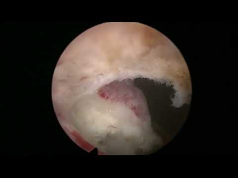
A sixth prostate operation after GreenLight vaporisation, prostatic artery embolisation, two TURPs and a further GreenLight procedure had not relieved the patient's urinary symptoms. The description identifies residual anterior apical tissue obstructing urine passage and reports its removal using an en-bloc approach. The account describes the operative finding and removal, without supplying long-term symptom follow-up.
Documented details and sources
Only details explicitly stated in the sources. Missing information does not mean it did not occur.
- En bloc
We found anterior apical residual BPH tissue that was blocking the passage of urine, and removed it with an en-bloc approach successfully.
- Redo surgery
RL HoLEP, Case 39: the sixth prostate operation
- Multiple previous procedures
a man who had undergone a GreenLight PVP, a prostatic artery embolization, two consecutive TURPs, and another GreenLight PVP
Source checked: 2026-10-06.
Available transcript · English
Automatic YouTube captions, not clinically reviewed. Names, technical terms and numbers may contain errors.
Selected spellings of enucleation, morcellation and holmium terms have been edited. This is not a full audio or clinical review; the original captions remain the source.
They may contain transcription or translation errors; check the explanation in the video.
- hi this is Fernando men Sanchez and this is an interesting case of a man who had five previous prostate operations he had a PVP then prosthetic artery embolization then he had two turps and then another PVP and he was complaining of all the constellations of urinary symptoms and what we thought by ultrasound is that this man still had until your apical residual tissue so we did this uh cystoscopy and you could see an ultrasound that there was still Anonymous tissue remaining mostly epically and anteriorly like you can see in this in this ultrasound so we decided to go for it and try to remove this this anterior tissue that was blocking the passage you can see that bladder neck is wide open but
- there's still tissue anteriorly so it took us you know less than 15 minutes to do this but it really changed the the symptoms of the patient who is now doing really well um and this is common many of many times I think the fear of going into the sphincter and damagingcontinence might produce these situations where anterior tissue uh is obstructive so we have to do a careful um careful delineation of of the Apex you see there's no clear transition between the sphincter and the anterior part so you know you have to just go for it try to try to understand you know where the edge of this thing could be sometimes you can see a little bit between the apical anterior tissue and the anterior part of the sphincter
- in this case there was no clue of where the limit would be so I did an initial careful um marking the white line as I as I call it and then as always when we do redo cases I try to explore how much um BPH tissue there is left circumferentially not only uh trying to remove whatever obviously remains but always trying to explore a little bit of the of the you know also the posterior tissue because sometimes we find that um there is there is residual tissue on the other hand and you can see there's a bump of tissue there that is probably BPH residual tissue which is amazing after so many ghosts to to the prostate so also here I was a little bit concerned about um you know a possible plan extra after
- anything we do so you'll see how things turned out at the end in this case I would think I was using the classic holmium the classic 100 watt um laser from luminous that uh we still have and we still use sometimes especially when you know when we are very busy and we work in two different operating rooms so we we might use the you know different different consoles for for different operating rooms so we don't have to keep moving stuff from one place to the next so here you see that I'm trying to look for orientation I'm trying to see how the tissue looks like when we come up words from the from the lateral plane trying to trying to understand so very carefully coming up you see coming up trying to follow the Contour and then of
- course going to the Apex to check uh what happens I think I was having some trouble with the fiber in this case so they probably would have taken 10 minutes if if we didn't have this Fiverr going in and out thing as you can see now here I'm putting the fiber in again I think yeah so there we are you know trying to understand trying to be careful anteriorly but of course we don't want to to leave more tissue no this man had already too many operations on his prostate and um yeah this is this is a not very common but this is a situation we see sometimes and uh you know if you do a cystoscopy you see the bladder neck open and maybe you don't you don't realize that what the bump you're seeing above is not just a
- sphincter but there is there is a residual BPH tissue there that is behaving obstructively so I think the transfectal ultrasound is a nice idea to to check in this cases and the if there is uh remaining BPH tissue which would be extremely easy after you know turp for example uh uh then it's it's it's worth going in and and giving it a go again and uh removing this this anterior tissue I thought it was an interesting uh case to show not very difficult but again it's uh we have a tool which is this laser we have uh uh anatomical knowledge and then we have some you know we have to face the cases that come you know this patient was was saying to me you know I have had five operations before it's it's
- absolutely crazy please help me because I cannot I cannot live like this you know and uh very frustrated actually he came from abroad and he had seen some of these videos and he was he was interested in this ultrasound evaluation again I had some trouble with the fiber so that was quite slow and I'm sorry for that um fiber in and out thing there there it is again and uh as you can see now we progress like as usual now the Apex has totally been released from the from the sphincter anteriorly and now it's a matter of going around going around and dissecting the the plane letting the laser do the the dissection carefully of course there is some fibrosity you know of the tissues after you know so many things done to this prostate but
- and the solution I found at the end uh to do this when I was meaning about preventing the bladder neck stricture was to to try to leave some of the bladder neck untouched so that hopefully it wouldn't it will not contract later on so you'll see that instead of a you know completely in Block operation I did uh you know half moon um Half Moon resection mostly of the anterior tissue preserving some of the some of the bladder neck and we we had a similar case of a patient who who was self-catheterizing because she he couldn't pee you know and he had also two two trps before and they told him you know the planet is wide open you have a weak bladder but we did a urodynamic testing and and he was clearly obstructed and and there was a
- bladder contraction so we we removed the tissue as well in the same fashion and he after four years of self-catheterization he was able to to void normally so for us it was a big big success and and the patient couldn't believe you know that because he had assumed that he had to self catheterize for life so interesting stuff I think we are learning a lot of anatomy with this uh and block technique and I think it's a very very useful uh technique and and the you know anatomic aspect of of it I think it's extremely attractive now trying to get rid of the problem once and for all so here again trying to follow the anterior line and all I always trust my lines a lot here I'm trying to remove that bump uh that
- we saw before typically and posteriorly but here again I will try to preserve a little bit of the bladder neck at six o'clock so that the chances of a stricture after this are are less and um 30 is trying to connect the lines and trying to connect the opening uh of the bladder neck in this side with the opening on the other side and there it goes I think that's the end of the and now we will have a look with translator ultrasound what I do is I keep the endoscope inside the bladder so the bladder is is filled of course I retract it so I can see the the fossa maybe keep the the endoscope in the in the urethra and at the same time we introduce a translator Sun probe to to have a look at things and how they look
- and I think it's a it's a very nice view of you know the difference pre and post-operatively of how the apical uh tissue looks I think there's still a sphincter there and the patient was perfectly continent so I I did a little bit of hemostasis to morcellate the tissue out and then we did the the ultrasound to check that the tissue had been removed completely there is that the that's the external sphincter and now there's no curtain you know coming down from from the so there's no interior tissue coming down uh here I was opening the bladder neck a little bit on that side maybe to get some whiteness of of the bladder neck yeah some hemostasis and then the modulation of the very small piece of
- tissue as usual which was extremely fast of course because there was only two or three grams of tissue and I promise it's going to end very fast so bear with me to to look at the ultrasound well this is a change of instrument uh face there we go that's the nephroscope coming in which was a little bit damaged I think because there was some funny Optical effect no it looks good and that's the most later blade and that's the bit of tissue we took out which is almost instantly more selected so I'll show you now the the trans rectal ultrasound post-operatively and typically we have a sterile she's covering the transect ultrasound you can see the Apex you can see the very Montana on this beautiful image
- that shows that all the apical tissue has been removed anteriorly here I think I was telling my nurse to change the view to the longitudinal View and there you can see how all the tissue that was there anteriorly has gone so I hope you enjoyed this short video all the best bye bye